Top 5 Sun-Induced Skin Lesions in Dogs
Alexander Werner Resnick, VMD, DACVD, Animal Dermatology Center
Epidermal pigmentation and the haircoat are highly effective at protecting skin from damage induced by ultraviolet light (UVL) radiation. Skin lesions associated with sunlight primarily occur in areas of greater exposure, most specifically on the sparsely haired dorsal muzzle and on the glabrous, often unpigmented, skin of the axillae, flank folds, pinnae and ventral abdomen. Additional risk factors for sun-induced (actinic) cutaneous lesions include residing in regions of higher elevation and/or increased sunlight, housing (eg, indoors vs outdoors), and individual and breed propensity toward sunbathing. UVA (320–400 nm) penetrates the skin deeper than does UVB (ie, with relatively short wavelengths; 290–320 nm) and is associated with the most skin damage. Melanin absorbs wavelengths most effectively in the UVA region of UVL.
1. Solar dermatitis
Solar dermatitis (ie, sunburn) is caused by direct UVL injury to skin cells, with severity related to duration and intensity of sun exposure; this is not the same as the thermal burn that can occur in darkly pigmented and/or dark-haired body regions exposed for extended lengths of time to strong sunlight. Canine solar dermatitis (Figure 1) frequently occurs on the dorsal muzzle (nasal) and ventrum (truncal), although any poorly pigmented or unpigmented skin with prolonged sun exposure is predisposed. Nasal lesions begin as erythema and scaling at the junction of the nasal planum and dorsal muzzle, expanding caudally as inflammation causes adjacent hair loss, exposing the skin to additional UVL. Subsequent scarring and postinflammatory depigmentation can further develop, and lesions can wax and wane, depending on seasonal sunlight fluctuations. Australian shepherds are considered at most risk for nasal solar dermatitis.

Nasal solar dermatitis in an intact male American Staffordshire terrier (10 years of age). Alopecia with erythema is extending caudally from a normal-appearing nasal planum. Erythema is also present on the rostral muzzle and eyelid margins.
A primary differential for canine nasal solar dermatitis is discoid lupus erythematosus (DLE), which causes depigmentation of the nasal planum first, followed by crusting. In contrast, solar dermatitis lesions typically begin on nonpigmented skin at the junction of the nasal planum and dorsal muzzle; the pigmented nasal planum itself is only affected later.
Canine truncal solar dermatitis can occur in breeds and individuals with light pigmentation and inclination toward sunbathing: specifically the Dalmatian, bull terrier, boxer, bulldog, basset hound, beagle, and American Staffordshire terrier. Depending on the dog’s tendency toward right or left lateral recumbency, the exposed side is more affected than the down (ie, sheltered) side. Initial lesions of truncal solar dermatitis consist of mild erythema with subtle lichenification and scaling of the axillae and flank regions (Figure 2). After repeated exposure, skin thickening and comedones can develop with accentuation of follicular ostia, leading to deep pyoderma and scarring.

Truncal solar dermatitis in the same patient as Figure 1. Note the accentuation of normal skin wrinkling (lichenification) with erythema and slight scaling in the axilla. Follicular ostia are prominent.
2. Hemangioma
Multiple forms of hemangioma (HA) have been reported, but of relevance here is UVL-induced HA. Chronic solar damage is a key cause of dermal HA in dogs with strong clinical association between sun exposure and tumor development on the skin of the ventrum. Sun-induced HA is more common than UVL-induced hemangiosarcoma (HSA) and may represent progression from vascular alteration to malignancy. In addition, UVL-induced HA can be distinguished from non–sun-induced HA by concurrent presence of actinic dermatitis; sun-induced HA commonly presents with multiple tumors.

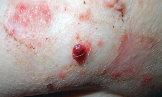
Hemangioma on the ventrum of a spayed American Staffordshire terrier (9 years of age).
Early lesions are punctate and appear as small, flat, purple discolorations. With time, lesions enlarge to well circumscribed, firm or fluctuant, red-to-purple plaques or nodules within the skin, varying in size from 0.5 to 4 cm (Figure 3). Larger lesions can ulcerate and bleed. As these tumors rarely metastasize, morbidity has been associated with excessive hemorrhage and secondary infection.

Spayed American Staffordshire terrier (9 years of age) with hemangiosarcoma lesion appearing as a crusted, deeply purpuric plaque on the ventral abdomen.
3. Hemangiosarcoma
UVL-induced HSA may represent malignant transformation within sun-induced HA or most often as progression from actinic keratoses (see below) and thus are most common on the ventral thorax and abdomen. As with HA, sun-induced HSAs are typically superficial in the dermis, often multiple in number. These tumors may be discrete or poorly circumscribed, dark red to purple, fluctuant, and often smaller than 2 cm in diameter (Figure 4). Prognosis is significantly better for sun-induced HSA than deeper and non–sun-induced cutaneous HSA, as the latter often can represent metastasis from distant sites of primary tumors.

Actinic keratoses on axilla of a spayed pointer crossbreed (9 years of age). Lesions are well demarcated, erythematous plaques with erosions, ulcers, and crusts.
4. Actinic keratosis
UVL can also cause direct damage to keratinocytes, producing mutations and resulting in expansion of mutated cell populations. The resultant actinic keratoses or sun-induced plaques that develop individually or in multiples are usually smaller than 1 cm in diameter. Early lesions resemble solar dermatitis, but the skin eventually becomes more palpably thickened and firm. Most lesions are crusted, and severe hyperkeratosis may resemble a cutaneous horn. In dogs, actinic plaques are most common on the glabrous and lightly pigmented skin of the axillae, flanks, ventral abdomen, and lateral areas of the extremities (Figure 5).
Actinic keratosis may represent a premalignant condition. Multiple studies have documented the close association of and genetic similarities between actinic keratosis and squamous cell carcinoma (SCC)1; actinic carcinoma in situ sometimes denotes similar pathology without dermal invasion by neoplastic cells.

SCC in the flank region of a spayed Dalmatian (5 years of age). The lesion is proliferative, ulcerated, and easily traumatized, leading to hemorrhage.
5. Squamous cell carcinoma
SCC is also noted to develop in areas of chronic inflammation, injury, and viral infection. Non–UVL-induced tumors can develop in different breeds and at different body locations than actinic disease. UVL-induced SCC rarely metastasizes. SCC is the second most common cutaneous malignancy in dogs. In humans, more than 80% of cutaneous SCCs may be associated with actinic keratosis.
While non–UVL-induced SCCs often present as single tumors, actinic-induced SCC may present as multiple tumors and associated with other lesions of solar dermatitis. SCCs appear as proliferative and ulcerated plaques (Figure 6). Older lesions may become crateriform and easily traumatized, leading to persistent serum drainage and/or hemorrhage.