Top 5 Postoperative Orthopedic Rehabilitation Considerations
Lisa Corti, DVM, DACVS, CCRP, North Shore Veterinary Surgery, Andover, Massachusetts, North Shore Community College, Danvers, Massachusetts
We sat down with Dr. Corti to further discuss rehabilitative exercises and how one size does not fit all when it comes to orthopedic rehabilitation. Listen to her episode of Clinician's Brief: The Podcast here.
Orthopedic surgery and postoperative rehabilitation can maximize functional outcomes in patients. Orthopedic repair can help prevent permanent loss of limb function, and a well-designed postoperative rehabilitation program can help prevent complications that could permanently affect quality of life.1,2 A multimodal approach involving surgical and rehabilitation teams can help achieve a successful outcome.1,3
Following are the author’s top 5 considerations during postoperative orthopedic rehabilitation.
1. Inflammation & Pain
Administration of NSAIDs, based on their well-researched efficacy, is currently the standard treatment for postoperative orthopedic inflammation and pain.4-7 The inflammatory phase of tissue healing typically occurs the first 3 to 5 days after injury8; however, edema, swelling, and pain from orthopedic surgery can last much longer. Thus, it is recommended that the duration of NSAID administration be determined on a case-by-case basis and NSAIDs be prescribed until suture removal when the patient can be reassessed and a decision made whether NSAID administration should be continued.5-7
In dogs and cats, a liposomal encapsulated bupivacaine formula infused into the surgical incision at the time of closure can help prevent proinflammatory cytokines from stimulating peripheral nociceptors, which may block transmission of pain for 72 hours.9 In humans, peripheral nerve blocks have been shown to provide better postoperative analgesia, promote earlier mobilization, and have a positive influence on surgical and rehabilitation outcomes.10,11 Peripheral nerve blocks can also improve a patient’s ability to tolerate manual rehabilitation therapies in the immediate postoperative period.11,12 Although peripheral nerve blocks have not yet been investigated for this purpose in veterinary medicine, the same benefit is likely to be seen in dogs and cats.
Cryotherapy uses cold temperatures to decrease inflammation and pain at the surgery site. Lowering tissue temperature can slow the metabolic rate of traumatized tissue, induce vasoconstriction, decrease sensory nerve conduction, decrease proinflammatory cytokine concentrations, and downregulate muscle excitability.3,13,14 The result is typically less inflammation and tissue damage, decreased edema and swelling, and reduced muscle spasms and pain levels.3,13 A cold pack made of a bag of ice or frozen vegetables wrapped in a thin towel or pillowcase, a commercial canvas ice pack (Figure 1), or a commercial cold pack that uses Velcro to fasten around the patient’s limbs and joints may be applied. The cold pack should be large enough to cover the entire surgical site, not just the incision; an application regimen of 10 to 30 minutes per session with an interval of 6 hours between sessions is recommended.3,13,14 Some cryotherapy devices can also apply compression, which improves contact between the cold source and the affected area on the patient. Improvements in pain scores, lameness scores, and stifle joint range of motion were noted when a pneumatic cold compression wrap was used around the stifle in dogs during the first 24 hours after tibial plateau-leveling osteotomy.3

Cryotherapy via a commercial canvas ice pack applied to the stifle of a dog. The ice pack is wrapped around the entire stifle, not just the lateral side. The contralateral limb is protected with a blanket.
2. Tissue Healing
Laser therapy (ie, photobiomodulation therapy [PBMT]) exposes tissue to electromagnetic radiation in a certain spectrum of light, leading to changes in electron and proton transfer that have biologic effects.15,16 These effects may include activation and production of growth factors, stimulation of cell growth and stem cell differentiation, promotion of vasodilation, angiogenesis, fibroblast proliferation, and epithelialization, with an overall acceleration in tissue healing.16,17 There is no standard dose or frequency recommended in veterinary medicine for PBMT. Doses using class 3B or class 4 lasers from 4 to 6 J/cm2 to 8 to 10 J/cm2 have been recommended by practitioners with experience in this field.15,18 In the acute postoperative period, daily administration of PBMT may be recommended with a greater interval between treatments as healing progresses.15,18
Massage has mechanical and physiologic effects that can aid in tissue healing during the postoperative period.19,20 Massage creates pressure differentials in which high-pressure areas increase venous and lymphatic outflow and low-pressure areas have an influx of new fluid. This flushing effect may enhance circulation, nutrient delivery, and waste removal and may decrease inflammation, pain, swelling, and edema.13,21 Deposition of scar tissue can also be minimized, as massage loosens muscles and tendons and enhances movement between tissue layers.20 A massage may be started immediately postoperation, with initial sessions performed with greater frequency and shorter duration. Several massage techniques exist, and there are many specialists certified in veterinary rehabilitation or massage.
Protected weight-bearing in the early stages after orthopedic surgery is an essential component of rehabilitation. In human medicine, no other technique has been shown to aid in the proper healing of injured bone, fibrous tissue, and muscle more than controlled physical activities.22 When a force or load is applied to connective tissue early after surgery, it causes increases in circulation and matrix synthesis that result in repair and remodeling of that tissue.22,23 Without the application of early controlled loading, the tissue would, at best, heal in a disorganized manner and, at worst, not heal at all.22-24 Dogs can be taken on a slow, controlled outdoor leash walk, with or without a sling or harness, or led to walk on an underwater treadmill. Cats can be encouraged to walk by placing trails of kibble on the floor, dividing meals into 3 to 4 separate food bowls placed around a room, or dragging a feather wand slowly across the floor. As tissue healing progresses and the patient improves clinically, the duration, frequency, and speed of walks or activity can be increased.24 It is generally safe to increase the amount of activity by 10% to 15% per week if the patient remains comfortable and the surgery site is not compromised.24
3. Joint Range of Motion
Passive range of motion (PROM) therapy involves movement of a joint with no muscle contraction and is commonly used in the early postoperative period to decrease pain and scar tissue formation, restore joint pliability, maintain flow and health of synovial fluid, and prevent muscle contracture.23,25,26 Patients are typically maintained in lateral recumbency with the affected limb pointed up. The joint to be manipulated is isolated, then passed slowly, gently, and continuously into flexion and extension while a range of motion that is comfortable for the patient is maintained.
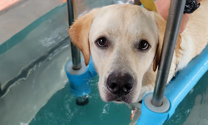
PROM therapy can be stopped once the patient is able to consistently bear weight on the affected limb and undergo active range of motion (AROM) therapy. Techniques that enhance AROM include assisted or independent leash walks, underwater treadmill therapy,27,28 and use of stairs and ramps29 and cavalletti rails (Figure 2).30 Swimming may improve AROM and, in postoperative cranial cruciate surgery patients, has been shown to result in greater range of motion in the stifle and tarsal joints as compared with walking31; however, this increase in range of motion is attributed to an increase in joint flexion.31 Because diminished joint extension is a common finding in both pre- and postoperative orthopedic patients, underwater treadmill therapy may be more effective in restoring joint extension than swimming.28 Underwater treadmill therapy can be tailored to the individual needs of the patient through alterations in water depth, belt speed, and activity duration and can begin as soon as the patient’s incision is healed.

A dog walking over cavalletti rails. The left thoracic limb and right pelvic limb are fully extended, whereas the right thoracic limb and left pelvic limb are hyperflexed. Navigating cavalletti rails is a versatile activity in which changes in distance between and the height of the rails boost AROM, gait retraining, and strength training. This dog is also engaging and strengthening core muscles.
4. Gait Retraining
In gait retraining (ie, neuromuscular rehabilitation), certain exercises are performed to stimulate reconnection between injured skeletal muscles and neurons. This typically results in limbs being retrained to function as they did prior to surgery.32,33 Exercise performed during muscle reinnervation stimulates the healing of injured and atrophied motor pathways, improves recovery of the neuromuscular response, restores neuromuscular feedback systems at peripheral and central levels, and accelerates recovery of the affected limb.32,33 These exercises are designed to challenge the patient’s balance and stimulate proprioception and may consist of the patient participating in underwater treadmill therapy; walking on and off different surfaces (eg, grass, pavement, dirt, leaves); navigating a makeshift obstacle course made of objects varying in height and length; walking through, over, and around cavalletti rails, caution cones, or tires; undergoing instability training on a wobble board; and/or standing on a physioroll (Figure 3). Additional exercises can include the therapist walking while holding the patient’s thoracic limbs in a “dance” position or walking while holding the patient’s pelvic limbs in a “wheelbarrow” position (Figure 4).

A dog being held with its thoracic limbs on an unstable physioroll. Simultaneous gait retraining and strength training occur while the patient also engages core muscles.


A therapist walks while holding a dog’s thoracic limbs in a “dancing” position (A) and with a dog’s pelvic limbs held in a “wheelbarrow” position (B). These exercises are used for gait retraining that also strengthens specific muscle groups in the limbs.
5. Strength Training
Strength training involves repetitive use of exercises that challenge muscles to contract against greater mechanical resistance.34,35 This should increase the patient’s muscle mass and strength and help the patient regain previous levels of muscle and limb function.34 Techniques used to increase the effort with which the limbs have to work may include decreasing the water depth in an underwater treadmill and increasing the speed at which the treadmill belt rotates.30 Similar techniques can be applied to land exercises: Increasing walking speed or jogging increases the impact on the force applied to limbs, and dogs can be trained to wear small leg weights; dogs can be encouraged to pull a sled with weights gradually added over time; poles can be raised during cavalletti training; walking pace can be increased during “dancing” and/or “wheelbarrowing”; and walking up and down steeper inclines can improve strength. Having the patient perform repetitive sit-to-stand exercises, which engage the hip and pelvic limb muscles, and down-to-stand exercises, which engage the thoracic and pelvic limbs, can strengthen specific muscle groups.
Strengthening Core Muscles
The need for strong abdominal and lower back muscles (ie, a strong core) is emphasized in humans, as a strong core improves the ability for physical activity.35,36 Although it may be more difficult, working on core strength in dogs and cats should not be overlooked. Having a patient stand on an unstable platform (eg, wobble boards, Bosu balls, physiorolls) can encourage engagement of the patient’s core muscles while the patient tries to maintain balance and avoid falling.30 The patient’s abdomen can be scratched or tickled while it stands on a moving wobble board to promote further contraction of the core muscles.37 Both dogs and cats can be trained to roll over on command. Rolling requires significant core strength; patients can be encouraged to repeat this activity during timed sessions.30 Patients can be made to stand with 2 paws up for a specified amount of time and a given frequency while the therapist holds a thoracic limb and the contralateral pelvic limb off the ground.37 Standing on 2 limbs in this manner stimulates contraction of the abdominal wall, back, and upper limbs and may aid in strengthening core muscles.
Conclusion
Postoperative physical rehabilitation is an important component in the treatment of surgical orthopedic conditions in dogs and cats. Although there is a need for more high-quality scientific research in veterinary medicine, strong evidence-based research in human medicine can be used for rehabilitative methods in dogs and cats. Methods that treat inflammation and pain, promote tissue healing, enhance joint range of motion, support gait retraining, and improve muscle strength should be considered for any postoperative orthopedic rehabilitation program.
Want to learn more?
AROM = active range of motion, PBMT = photobiomodulation therapy, PROM = passive range of motion