Top 5 Ocular Complications of Brachycephaly in Dogs
Noelle McNabb, DVM, DACVO, Animal Vision Care & Surgical Center, Studio City, California
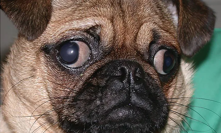
Shallow bony orbits and exophthalmos in a 2-year-old pug. Marked macroblepharia and lagophthalmos are also present.
In brachycephalic dogs, the skull is disproportionately shorter and wider than is typical for other dog breeds. The muzzle is flattened, and the nose and maxilla are markedly shortened. This central facial flattening results in poor medial canthal support and folds of nasal skin, which causes secondary entropion, trichiasis, and pigmentary keratitis. Bony orbits are shallow, which causes exophthalmos. Globe protection is often further diminished by macroblepharia (ie, overly large palpebral fissures caused by excessive horizontal eyelid length).1
Chronic exposure and desiccation of the ocular surface can result in exposure keratitis and tear film deficiencies, which are often compounded by concurrent keratoconjunctivitis sicca (KCS).2,3 These associated disorders of the eyelids, cornea, conjunctiva, and nasolacrimal system are known collectively as brachycephalic ocular syndrome.2
Even minor facial injuries can have devastating ocular consequences for brachycephalic dogs. Because ocular complications of brachycephaly can threaten vision, daily therapeutic and prophylactic management is often necessary to maintain long-term vision and comfort.
1. Lagophthalmos
Lagophthalmos is the inability to completely close the eyelids (Figure 1). Impaired lid closure in brachycephalic dogs is attributed in part to their shallow bony orbits, resulting in exophthalmos. Lagophthalmos is often exacerbated by concurrent macroblepharia.1-3 Commonly affected breeds include the shih tzu, pug, English bulldog, Chinese shar-pei, Pekingese, Boston terrier, and Lhasa apso.

Shallow bony orbits and exophthalmos in a 2-year-old pug. Marked macroblepharia and lagophthalmos are also present.
Lagophthalmos results in chronic ocular surface desiccation, which leads to exposure keratopathy (ie, noninflammatory irritation of the cornea). Lagophthalmos is characterized by corneal opacification, related to progressive epithelial fibrosis and vascularization. Lagophthalmos also contributes to tear film deficiency (ie, increased evaporation of the aqueous portion of the trilaminar tear film), which further devitalizes the corneal epithelium, predisposing it to ulceration, collagenolysis, and infection.
Lagophthalmos benefits from daily treatment with lacrimomimetics (ie, tear substitutes). Several effective lacrimomimetics are available over the counter. These products typically contain dextran, cellulose, polyvinyl alcohol, polyethylene glycol, or viscoelastic substances (sodium hyaluronate or chondroitin sulfate). Products containing hyaluronic acid are particularly efficacious.4
Animals with pronounced macroblepharia can benefit from medial or lateral canthoplasty, a procedure that reduces the size of the palpebral fissure, improving blink quality and globe protection and potentially reducing corneal trauma.2,3 Tear film distribution and quality also increases, improving corneal epithelial health and transparency.
2. Tear Film Deficiency
Dry eye is a disorder of the tear film caused by tear deficiency or excessive evaporation. KCS (ie, aqueous tear deficiency) occurs with increased frequency in brachycephalic dog breeds, including the English bulldog, pug, Lhasa apso, Cavalier King Charles spaniel, and shih tzu. KCS is a local autoimmune-mediated inflammatory condition affecting the lacrimal and nictitans glands.5 Complications from KCS include mucopurulent ocular discharge, conjunctivitis, corneal ulceration and infection, collagenolysis, and corneal perforation. Chronic disease incites progressive corneal fibrosis, vascularization, pigmentation, and vision loss.

Medial canthal entropion and caruncular trichiasis in an 8-year-old shih tzu with chronic KCS. This combination of disorders has resulted in diffuse pigmentary keratitis and blindness.
Lagophthalmos increases tear evaporation and impairs tear distribution, resulting in a thin precorneal tear film. The development of concurrent KCS substantially increases the incidence and severity of ulcerative keratitis, pigmentary keratitis, and subsequent vision loss (Figure 2).
Diminished corneal sensitivity further compromises corneal health. Reduced corneal innervation has been demonstrated in brachycephalic dogs, and nerve density is lowest at the center.6 Diminished corneal sensation decreases protective lacrimation that would otherwise aid in washing away fine debris, irritants, and abrasive particles from the surface of the cornea.
Brachycephalic patients with Schirmer tear test values <12 to 13 mm/minute and concurrent clinical features of KCS benefit from medical therapy. Topical cyclosporine is effective in stimulating tear production. Cyclosporine 1% or 2% benefits patients with KCS through selective T-helper lymphocyte suppression and direct lacrimostimulation. If cyclosporine therapy fails in a patient with KCS or the patient is sensitive to cyclosporine or the lipid bases in which it is formulated, tacrolimus is a good alternative. Tacrolimus 0.02% administered topically q12h has been shown to be effective in increasing tear production in both patients that received no previous tear stimulation therapy and in those that responded insufficiently to cyclosporine.7

Nasal fold trichiasis in 3-year-old pug. Note the prominent nasal fold and medial canthal entropion with secondary pigmentary keratitis.
3. Medial Canthal Entropion & Trichiasis
Poor medial canthal support in brachycephalic dogs often results in entropion at the medial canthus and ventromedial aspect of the lower eyelid. Entropion and trichiasis (Figure 3) adversely affect the corneoconjunctival surface, contributing to epithelial ulceration, fibrosis, vascularization, and pigmentation.
In affected dogs, medial canthoplasty can eliminate chronic corneal friction and vision-threatening keratitis.2 This procedure can be combined with nasal-fold–shortening procedures in dogs with concurrent nasal fold trichiasis.1,2

Medial canthal and ventral medial entropion in a 5-year-old pug. Secondary corneal and conjunctival melanosis are also present.
4. Pigmentary Keratitis
Chronic keratitis can develop secondary to the oculofacial conditions associated with brachycephalic ocular syndrome. The resultant corneal vascularization and pigment deposition typically begin at the medial limbus and extend laterally, leading to progressive visual impairment (Figure 4). Coexisting eyelash disorders such as distichiasis (ie, an abnormality in which aberrant cilia emerge from the meibomian gland orifices) and ectopic cilia (ie, cilia that protrude through the palpebral conjunctiva) can contribute to keratitis and are prevalent in some brachycephalic dog breeds, including the shih tzu, pug, and English bulldog.2
KCS also contributes to the development of pigmentary keratitis. Pigmentary keratitis occurs with higher incidence in brachycephalic dogs, particularly those with KCS and lagophthalmos. These patients typically experience diffuse corneal pigmentation and blindness because increased desiccation and persistence of corneal vascularization allow more pigment to enter.
The pug is at particular risk for developing pigmentary keratopathy, and a genetic component has been suggested.8
5. Ocular Trauma
Brachycephalic breed dogs are at greater risk for ocular trauma than dogs of other breeds because of increased globe exposure. The shallow bony orbits provide considerably less globe protection as compared with their dolichocephalic and mesocephalic counterparts.<sup1-3 sup> Protection is further impaired by lagophthalmos. Injury rates are especially high in dogs <5 months of age because they have not yet developed a menace response. In brachycephalic dogs of any age, globe proptosis can result from trauma that occurs when jumping off furniture, when playing with other pets, or during routine bathing and grooming.
Decreased central corneal sensitivity in brachycephalic dogs enables potentially irritating particles and debris to linger on the corneal surface, causing epithelial damage and erosion. These microabrasions are susceptible to infection and may result in collagenolysis, perforation, and vision loss.
Urgent medical treatment of ocular injuries maximizes success in preserving vision. The anatomic abnormalities and tear film deficiencies present in brachycephalic dog breeds result in a higher incidence of ocular trauma and more severe forms of ocular injury, and normal healing processes are compromised by these abnormalities.
Conclusion
Diligent daily monitoring of the eyes of brachycephalic dogs is crucial for identification of ophthalmic disease and ocular injury and for prompt vision-preserving medical and/or surgical intervention.
KCS = keratoconjunctivitis sicca