Top 5 Glaucoma Drugs
Erin M. Scott, VMD, University of Wisconsin–Madison
Gillian J. McLellan, BVMS, PhD, DVOphthal, DECVO, DACVO, MRCVS, University of Wisconsin–Madison
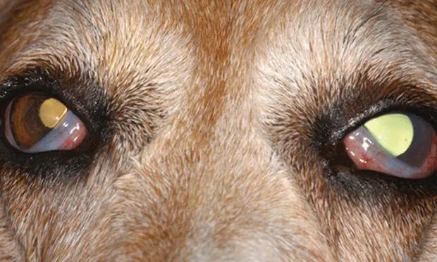
Glaucoma is typically associated with elevated intraocular pressure (IOP) resulting from impaired aqueous humor outflow and leads to irreversible damage to the optic nerve and retina.1,2 Primary glaucomas are relatively common and have strong breed predilections in dogs but are rarely diagnosed in cats.1,3,4 Secondary glaucomas are as frequent as primary glaucomas in dogs and constitute 95% to 98% of glaucoma cases in cats.1,4-7 They are usually secondary to an ocular or systemic disorder that impairs aqueous humor outflow (eg, uveitis, neoplasia, lens luxation, trauma).8 Since glaucomas are heterogeneous in terms of pathogenesis, therapy must be tailored to each patient (see table Commonly Used Glaucoma Drugs at a Glance). Glaucoma medications target IOP by either decreasing production or increasing outflow of aqueous humor.
TOP 5 GLAUCOMA DRUGS
Carbonic anhydrase inhibitors
Prostaglandin analogs
β-blockers
Miotics
Hyperosmotic agents
1. Carbonic Anhydrase Inhibitors
Carbonic anhydrase, an enzyme present in the ciliary body epithelium, contributes to aqueous humor production. Carbonic anhydrase inhibitors (CAIs) can decrease aqueous humor production in dogs and cats by over 40%.9,10 Dorzolamide 2% effectively lowers IOP when administered q8h.10-13 It may be used in the management of acute glaucomatous crises and for long-term control of IOP. Dorzolamide may be considered a safe first-line drug of choice for dogs and cats with glaucoma, regardless of cause. It is often used in combination with other glaucoma drugs, particularly in primary glaucoma patients. Topically administered CAIs can cause transient conjunctival irritation or more persistent conjunctivitis in cats and dogs; they may be associated with vomiting, hypersalivation, and inappetence in cats.4 Patients intolerant of dorzolamide may show fewer adverse effects with brinzolamide. A fixed combination of 2% dorzolamide and 0.5% timolol is available and, when administered q12h, has been shown to be more effective than timolol or dorzolamide alone at reducing IOP in glaucomatous dogs.14
2. Prostaglandin Analogs
Prostaglandin analogs decrease IOP in humans by increasing the outflow of aqueous humor through the uveoscleral pathway.15 Because of species differences in the prostanoid receptors in the iris sphincter muscle, commercially available prosta-glandin analogs cause an intense miosis in both dogs and cats but not in humans.16 This miosis may mechanically reverse some forms of pupillary block (ie, an obstruction to the flow of aqueous humor between the pupillary margin and the anterior lens capsule), which is a proposed mechanism of acute IOP elevation in dogs with primary angle-closure glaucoma.1 Topical latanoprost 0.005%, applied q12–24h, is rapid, potent, and effective in decreasing IOP in glaucomatous dogs by 65% to 70%.17 Although latanoprost transiently lowers IOP in cats with rare, primary congenital glaucoma, it may increase IOP in some feline glaucomas.4,18,19 Prostaglandin analogs, such as latanoprost and travoprost, are important in emergency and long-term treatment of primary glaucoma in dogs but should be used cautiously in feline glaucoma, as they are less likely to effectively lower IOP and may exacerbate underlying uveitis or dramatically increase IOP. Use of latanoprost is often contraindicated in secondary glaucoma, particularly with preexisting uveitis, lens luxation, and/or vitreal prolapse. Caution should be observed if there is doubt whether the glaucoma is primary or secondary. Adverse effects in dogs include ocular inflammation and visually impairing miosis.
3. β-Blockers
β-blockers decrease IOP by reducing blood flow to the ciliary body, thereby decreasing aqueous humor production. β-blockers have limited use as a single agent since they only modestly lower IOP in glaucomatous dogs and normal cats. While the β-blocker, timolol, may have significant additive effects when combined with CAIs in dogs, this additive effect was not seen in normal cats.14,20,21 β-blockers, applied topically q12h, are not effective during sleep, when sympathetic tone is lower but significant IOP increases may occur in glaucoma patients.22,23 However, β-blockers can play an important role in glaucoma prophylaxis in dogs. Similar to the miotic drug demecarium bromide (see Section 4), betaxolol given q12h significantly prolonged onset of primary glaucoma in the fellow eye of dogs.24 Adverse effects may include bradycardia, arrhythmias, and bronchoconstriction, and β-blockers may be contraindicated in cats with respiratory conditions.
4. Miotics
Miotic agents increase aqueous outflow by constricting the pupil and contracting the ciliary muscle. Their use effectively and modestly reduces IOP in most canine and feline glaucomas, except for those associated with uveitis or lens luxation.2 Demecarium bromide 0.125% to 0.25%, administered q12–24h, is a long-acting anticholinesterase that can be safely administered for prolonged periods in dogs.25 Currently, demecarium bromide is not available commercially and must be compounded for ocular use. The direct acting cholinergic miotic, pilocarpine, is less efficacious and is poorly tolerated by veterinary patients because of its propensity to cause ocular irritation and inflammation. Hence, pilocarpine is infrequently used in the management of canine or feline glaucomas.
Adverse effects include ocular irritation and exacerbation of intraocular inflammation. Similar to prostaglandin analogs, miotics should only be used with extreme caution in patients with preexisting uveitis or a tendency to pupillary block (eg, associated with lens luxation or vitreous prolapse). Prophylactic administration of demecarium bromide in conjunction with a topical corticosteroid significantly prolongs the time to onset of IOP elevation in the fellow eye of dogs with primary angle-closure glaucoma, from 8 months (in untreated dogs) to 32 months.24
5. Hyperosmotic Agents
Mannitol is an important systemic agent for decreasing IOP in severe and refractory glaucoma and should be used in emergency cases only. Mannitol increases the osmotic concentration of blood perfusing the eye, causing a marked reduction in aqueous humor production and vitreous volume.1 Vitreal “dehydration” directly decreases IOP and allows the intact lens to move posteriorly, increasing outflow of aqueous humor and reducing pupillary block.1
Mannitol 20% is administered IV (1–2 g/kg) over 15 to 20 minutes. A dramatic IOP reduction typically occurs within 30 to 60 minutes and lasts for 5 to 6 hours.1 Mannitol should be gently warmed and given through an administration set with a filter, as concentrations >15% may crystalize at low temperatures. Profuse diuresis is to be expected but water intake must be controlled. Mannitol is contraindicated in patients with chronic renal failure and congestive heart failure, and may be of limited efficacy in patients with breakdown of the blood-aqueous barrier (eg, from uveitis or trauma). After IOP reduction, ocular perfusion is restored, and topical glaucoma medications, which should be administered concurrently with mannitol, are rendered more effective. The need for IV mannitol has been reduced and the practice of dispensing the orally administered hyperosmotic, glycerol, has been largely superseded by topical prostaglandin analogs (eg, latanoprost), which have been proven effective in the emergency management of acute glaucomatous crises.
Final Thoughts
Glaucoma therapy should aim to lower IOP, preserve vision, and eliminate pain. A delicate balance between aqueous humor production and outflow must be achieved, and IOP should be frequently monitored. Medical and surgical therapies are often required to maintain a comfortable, visual eye with glaucoma. Vision should be carefully evaluated, as an irreversibly blind and painful eye does not warrant aggressive medical therapy. Evaluating the risk for glaucoma in the fellow eye and managing accordingly are imperative. The decision to treat medically, surgically, or both depends on patient vision and temperament, owner ability to treat, and associated costs, as well as the underlying cause of glaucoma.1 When possible, the advice of a veterinary ophthalmologist should be sought.