Top 5 GI Parasites in Companion Animal Practice
Chris Adolph, DVM, MS, DACVM (Parasitology), Zoetis Animal Health
Images courtesy of the National Center for Veterinary Parasitology.
GI parasites are common pathogens that companion animal veterinarians must address. One study estimates that as many as 62% of pet owners allow dogs and cats on their beds.1 As the relationship between pets and owners continues to evolve, it becomes even more important for veterinarians to discuss parasitism, both internal and external, with every pet owner at every visit. To have an informed discussion on common parasites, veterinary team members need to understand prevalence, life cycle, pathogenesis, diagnostic challenges, and control measures. This article discusses the 5 most commonly detected intestinal parasites, in this author’s opinion, in small animal practices.
1. Hookworms
Common hookworm species encountered in companion animal practice include Ancylostoma caninum, A braziliense, Uncinaria stenocephala (dogs), and A tubaeforme (cats). Hookworm life cycles are mostly similar; minor differences exist. For example, A tubaeforme in cats may utilize a rodent paratenic host to complete the life cycle.
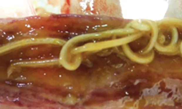
The definitive host may acquire a hookworm infection via ingestion of environmental third-stage larvae (L3), through the transmammary route (dogs), or by predation of infected paratenic hosts.2 Method of acquisition and age of the definitive host will influence the prepatent period and pathogenicity. Hookworm larvae are also capable of migrating to peripheral tissues, where the larvae can survive for years and develop into adults (Figure 1).3 This can explain why some pets remain positive on fecal flotation after anthelmintic therapy.

Adult Ancylostoma spp infection in a dog.
Clinical signs of hookworm infection can include anemia, melena, and occult blood in the feces. Shock and death can be seen in untreated puppies of nursing age with heavy infections.4 Coughing, nasal discharge, fever, and signs of pneumonia are possible during larval migration through the lungs.2
Diagnosing hookworm infection is usually done by fecal flotation. Both Ancylostoma spp and U stenocephala eggs are recognized by their elliptical shape, smooth shell wall, and grape-like clusters of cells called morulae. Ancylostoma spp eggs are slightly smaller than U stenocephala eggs.5 However, lack of detection does not rule out infection.
Hookworms are zoonotic, although feces are not immediately infective. Humans acquire hookworms when unprotected skin (eg, feet, buttocks, hands) is exposed to ground where larvae have reached the infective L3 stage. The invading larvae cause a dermal hypersensitivity reaction. Hookworms rarely reach adulthood in humans, although cases have been reported.6
2. Roundworms
Like hookworms, ascarids comprise multiple species that infect dogs and cats. Toxocaracanis (dogs) and T cati (cats) are the most common ascarids found in their respective definitive hosts. T leonina can be found in both dogs and cats, whereas Baylisascaris spp, typically a parasite of raccoons, can infect dogs.
The definitive host passes noninfective (unembryonated) eggs into the environment where, after time, the eggs embryonate and become infective not only to their definitive hosts (dogs and cats) but also to a range of other paratenic hosts, including humans.7 Dogs and cats can acquire ascarids by ingesting infective eggs. In this case, the infective larvae penetrate the intestinal mucosa; enter the hepatic portal system; migrate to the lungs, trachea, and pharynx; and are swallowed and complete their development in the GI tract (Figure 2).8 Dogs and cats may also become infected by ingesting paratenic hosts. A wide range of animals may serve this role, including rodents, rabbits, farm animals, and earthworms.7 When the paratenic host route is taken, the above-mentioned tracheal migration does not occur; instead, ingested larvae develop fully within the GI tract.8T canis, but not T cati, can be passed transplacentally from an infected female to her puppies. The source of infection can be activation of latent larvae in the female’s tissue or from a recent infection.9

Adult Toxocara canis infection in a dog.
Roundworm infections, particularly minor burdens, are usually subclinical. Young animals often have abdominal distention, intermittent diarrhea, and a thickened intestine. Death can occur in young animals with heavy infections as a result of possible hepatic and pulmonary damage by migrating larvae. Pneumonia associated with worm migration in prenatally infected puppies can be a major cause of mortality.7 Although pneumonia is minimal with T cati infection, migration of this nematode produces a medial hypertrophic lesion of the pulmonary arterioles that is also produced when cats are infected with Dirofilaria immitis and Aelurostrongylus abstrusus.
Diagnosing patent roundworm infection is based on recovery of eggs, which are characterized by a round, single-celled embryo within a thick shell wall. T canis is slightly larger than T cati.5 As with hookworms, lack of detection does not rule out infection.
Ascarids are zoonotic and, like hookworms, their eggs are not immediately infective. Unlike hookworms, however, ascarid larvae will cause damage deeper than the dermis. Aberrant migration of larvae can cause damage to the liver (visceral larva migrans), eyes (ocular larva migrans), and CNS (neural larva migrans).
3. Whipworms
Dogs infected with Trichuris vulpis pass single-celled eggs that are not infective initially. An infective first-stage larva develops while the eggs are in the environment, but they will not hatch unless swallowed by the definitive host.3 Once the eggs are ingested, development to adult worms is limited to the intestine with no larval migration outside the intestinal tract. (Figure 3).2

Adult Trichuris vulpis infection in a dog.
Dogs infected with whipworms can remain free of clinical signs; however, heavy infections cause episodes of diarrhea (sometimes bloody) alternating with periods of normal stools. It is not uncommon for owners to delay care because of the intermittent nature of this infection.
Diagnosis is best achieved with centrifugal flotation rather than passive flotation because the T vulpis egg is much heavier than other nematode eggs. Eggs are recognized by their barrel shape, bipolar plugs, and smooth shell wall surface.5 It is important to distinguish between Trichuris spp and Eucoleus spp eggs, as both possess bipolar plugs.
The zoonotic risk with T vulpis is very limited.
4. Giardiasis
Multiple species and assemblages of Giardia spp infect a wide range of hosts. Transmission of Giardia spp is via the fecal–oral route. Cysts are passed in feces and are capable of contaminating food, water, and fomites. Once cysts are ingested, excystation results in trophozoite emergence and attachment to the small intestinal epithelial cells, potentially causing inflammation and mucus production. This can lead to a maldigestive or malabsorptive enteritis. Trophozoites usually form cysts that are infective to the next host and are passed in feces. However, hosts can auto-infect by ingesting passed cysts while grooming the perineal area.
Signs of Giardia spp infection can be subclinical, acute, or chronic. Dogs and cats can be presented with pale, pasty, foul-smelling stools as well as vomiting. Diarrhea can begin as soon as 5 days after exposure.10
Diagnosis is by detection of antigen in feces or detection of trophozoites or cysts in stool. For cysts, centrifugation with zinc sulfate solution is considered the most sensitive. Occasionally trophozoites can be detected on a direct smear (Figure 4). The zoonotic potential with Giardia spp is low.

Giardia spp trophozoites from a dog (magnification 400×).
5. Coccidiosis
Enteric coccidia encountered in small animal practice include Cystoisospora canis, C ohioensis, C burrowsi, and C neorivolta in dogs and C felis and C rivolta in cats. Sporozoites, which are the infective stage, are contained in sporulated oocysts in the environment. Once a host ingests the sporozoite, it excysts from the sporulated oocyst and invades intestinal epithelial cells where it can evade the host immune response. Several rounds of sexual and asexual reproduction culminate with the production of oocysts. Pathologic changes result when developing stages emerge from the host epithelial cells. This results in GI bacteria colonizing the ruptured enterocyte.3 Enteric coccidia are very host-specific. Dogs and cats are capable of ingesting oocysts of other host species (other Cystoisospora spp or Eimeria spp). However, these coccidia simply pass through the animal and are of no pathologic significance; no treatment is required.
Chronic diarrhea is the main finding in clinical coccidiosis. Most clinical cases occur in puppies and kittens that undergo a stressor, such as weaning, change of home, or concurrent illness. Fatalities may occur during the early asexual stages of reproduction before oocysts develop. It is important to be aware of other causes of diarrhea in older patients in which oocysts are detected.
Diagnosis is by a combination of history, clinical signs, fecal flotation, and exclusion of other potential causes. On flotation, oocysts can be unsporulated, containing a single round cell, or sporulated, containing 2 sporocysts that each contain 4 sporozoites (Figure 5).5 Oocysts may be found in clinically normal puppies and kittens, and care must be taken not to over-interpret their relevance. Finding coccidia in a diarrheic puppy or kitten does not necessarily mean that coccidiosis is the cause of disease; further investigation should occur.

Cystoisospora ohioensis sporulated oocysts from a dog (magnification 400×).
Closing Thoughts
Depending on geographic location, the prevalence of these and other parasites varies. Maps by the Companion Animal Parasite Council (CAPC), which provide a county-by-county breakdown of prevalence estimates for common intestinal parasites, can be a valuable resource for veterinarians in getting a better idea of what is being diagnosed in their area. If prevalence in a practice is less than is usually reported in the area, the practice should evaluate its diagnostic methods and make improvements as needed. When armed with current information, most owners will be open to recommendations for keeping their pet free of parasites.
And the Most Common GI Parasite You Can’t Find…
The common tapeworms of companion animals in North America, Dipylidiumcaninum and Taenia spp, are found where intermediate hosts (fleas for D caninum; rabbits, sheep, or rodents for Taenia spp) exist.11 However, determining true prevalence without necropsy is impossible because of a lack of diagnostic stages in samples tested in practice. Because these tapeworms only shed proglottids intermittently, the probability of obtaining a proglottid is low. Likewise, if a proglottid is collected with a stool sample, processing has to allow the proglottid to rupture in order to release the embryos.

Adult Taenia spp in a dog.
Next, the flotation solution has to have a high enough specific gravity to float the embryo to the top. Failure at any step yields a false-negative result if the animal is truly infected. Therefore, fecal flotation and proglottid recovery are poorly sensitive tests for diagnosing tapeworms, but currently these are the only tests available to practicing veterinarians.
Recent studies show that more than 50% of dogs and cats at shelters were positive for tapeworms at necropsy. Yet fecal flotation failed to identify 100% of D caninum–positive cats, 94% of D caninum–positive dogs, 74% of T taeniaeformis–positive cats, and 43% of Taenia spp–positive dogs.12,13
Adult D caninum in a dog.
Many dogs and cats thus leave the veterinarian with undiagnosed tapeworm infections and no treatment. But are tapeworms really benign? A recent report highlighted a cat that required an enterotomy to resolve a tapeworm impaction.14
Until diagnostic methods improve, veterinarians should consider tapeworm infection in patients with past or current flea infestations, patients that hunt, and patients that roam. In addition, Echinococcus spp tapeworms are zoonotic, causing hydatid cysts in humans.
Editor's note: At the time of publication, Dr. Chris Adolph is affiliated with Zoetis Animal Health.