Top 5 Conditions Affecting the Pinnae
Andrew Rosenberg, DVM, DACVD, Animal Dermatology & Allergy Specialists, White Plains, New York, and Riverdale, New Jersey
The pinnal margins and pinnae of dogs and cats can be affected by many dermatologic diseases and disorders. Following are the author’s 5 most common conditions that affect only the pinnae, are most severe on the pinnae, or affect the pinnae prior to affecting other regions of the body. Infectious, ectoparasitic, immune-mediated, and neoplastic diseases and keratinization disorders should be considered as potential causes of pinnal diseases.
1. Pinnal Vasculitis
Vasculitis most commonly affects the pinnal margins of dogs and cats. Clinical signs can include alopecia, scaling, erosion, ulceration, crusting, and necrosis (Figure 1). In some cases, tissue loss may cause changes to the shape of the pinnae. Vasculitis is a histopathologic reaction pattern that signals the presence of inflammatory cells in blood vessels and is an immunologic response (ie, type III hypersensitivity disorder) that results in damage to vascular components of the dermis or subcutaneous tissue. Clinical signs can result when adequate oxygenation of tissue does not occur and blood vessels are damaged. A diagnosis of vasculitis is typically obtained through biopsy, and the underlying trigger should be determined. Numerous inciting agents have been implicated as factors that cause vasculitis, including drugs, vaccines (most commonly, rabies vaccine), food hypersensitivity, insect bites, malignancies, and infectious organisms. Many cases of vasculitis are idiopathic.1-3

Canine pinnal vasculitis. Pinnal margin alopecia, scaling, erosion, ulceration, and crusting can be observed.
Treatment of vasculitis depends on the severity of the case and the underlying cause. If an inciting cause can be determined, it should be removed and avoided in the future. Any underlying infectious disease that acts as an inciting cause should be treated (eg, tick-borne diseases can be treated with doxycycline). Immunomodulatory medications can be used to treat vasculitis. Pentoxifylline is often the treatment of choice. Pentoxifylline increases erythrocyte flexibility and decreases blood viscosity, thereby allowing increased oxygenation of damaged tissue. For patients that are nonresponsive to pentoxifylline, strong immunosuppressive medications (eg, glucocorticoids, cyclosporine, azathioprine) may be required. Some severe subforms of vasculitis, namely thrombovascular necrosis of the pinnae (Figure 2), may not respond to medication and may require surgical intervention. A partial pinnectomy can be performed, ideally with a CO2 laser, with care taken to remove all affected tissue.2

Thrombovascular necrosis of the pinna. Loss of tissue and necrosis of part of the pinnal margin can be observed.
2. Ceruminous Cystadenomatosis in Cats
Ceruminous cystadenomatosis is a disease that affects the concave pinnae, external orifices, and/or, occasionally, ear canals of cats. The cause of ceruminous cystadenomatosis is unknown; however, the formation of cystic structures has been suggested to be related to, although not generally associated with, otitis externa.4 Typical lesions include macules, papules, and/or vesicles and are multifocal-to-coalescing, gray-blue, and filled with a yellow, honey-colored fluid that can be expressed when the lesion is punctured (Figure 3). These cysts can be left untreated in mild cases, but severe cases can cause an obstruction that can lead to ear disease, predisposing the affected cat to chronic and recurrent otitis externa.5
The preferred treatment in advanced cases, particularly those causing recurrent otitis externa, is laser ablation with a CO2 laser; other medical management options are not effective. Otitis typically resolves once cysts are ablated; however, cysts can occasionally recur or the cat may develop new lesions.

Multifocal-to-coalescing, gray-blue macules, papules, and vesicles partially occluding the external ear canal caused by ceruminous cystadenomatosis
3. Scabies (Sarcoptic Mange) in Dogs
Canine scabies (ie, sarcoptic mange) is caused by the mite Sarcoptes scabiei subsp canis and may cause severe, nonseasonal pruritus in affected dogs. Scabies is spread through contact with other dogs or wildlife (eg, foxes, fox dens). In most cases, scabies affects the pinnal margins, with the pinnae being the first affected areas of the body.6 Other commonly affected areas include elbows and hocks. Lesions on the pinnal margins are typically scaly, crusted, alopecic, and erythematous (Figures 4 and 5). Most dogs infected with scabies exhibit the pinnal–pedal response (ie, kicking of the pelvic limb in response to the pinnal margin being rubbed).7
Although a canine scabies diagnosis is typically made through superficial skin scrapings, it can be difficult to find mites, even when they are present and lesions are apparent. An acaricidal trial is recommended if no mites are found but scabies is still suspected; avermectins can be an effective treatment option. New acaricides in the isoxazoline class of parasiticides (eg, sarolaner, fluralaner, afoxolaner, lotilaner) are the current treatment of choice but are extra-label and not approved for scabies treatment. Drugs in the isoxazoline class act through selective inhibition of arthropod γ-aminobutyric acid and L-glutamate–gated chloride channels.8-10 Medication should be administered according to normal recommendations for flea and tick control. All dogs in the household should be treated, and affected bedding should be washed in hot water or destroyed. Adjunct therapy with antihistamines, corticosteroids, or oclacitinib may be helpful in reducing pruritus associated with scabies, which is caused by a hypersensitivity reaction. Humans in the household exhibiting symptoms should seek advice from their physician.

Scaly and crusted alopecia caused by canine scabies

Bilateral scaly and crusted alopecia caused by canine scabies
4. Ear Margin Seborrhea
Ear margin seborrhea is a localized form of seborrhea that affects the pinnal margins in dogs. Dachshunds are predisposed to ear margin seborrhea, but it can also occur in other breeds (typically those with long, pendulous ears). Lesions start as scaly and adherent greasy casts that can progress and affect the entire ear margin (Figure 6). In severe cases, thick crusting with seborrheic debris and fissures can occur. Crusting and fissures can be painful and may result in shaking of the head and scratching, which can cause additional fissures and hemorrhage. Differential diagnoses include scabies, vasculitis, and sebaceous adenitis.
Ear margin seborrhea is not curable but can be controlled with various well-tolerated medications. Seborrheic shampoos that contain benzoyl peroxide, sulfur–salicylic acid, and/or phytosphingosine or ceramides are recommended. Affected areas can be bathed with these shampoos daily until resolution, then at a maintenance frequency. Weekly use of a topical blend of essential fatty acids may also be effective. It has been recommended that affected dogs not be allowed to sleep near forced-air heating ducts. Glucocorticoids may be administered if severe inflammation is present.11

Scaly and crusted hyperkeratotic adherent debris caused by ear margin seborrhea
5. Sebaceous Adenitis
Sebaceous adenitis is an inflammatory disease of the sebaceous glands that causes destruction and loss of these glands. Although the cause and pathogenesis of the disease are not known, breed predisposition and occurrence in family lines suggest a possible genetic component. Sebaceous adenitis can occur in any breed but is seen most often in standard poodles, Akitas, Samoyeds, vizslas, and poodle crossbreeds.12-14 Cats are rarely affected.
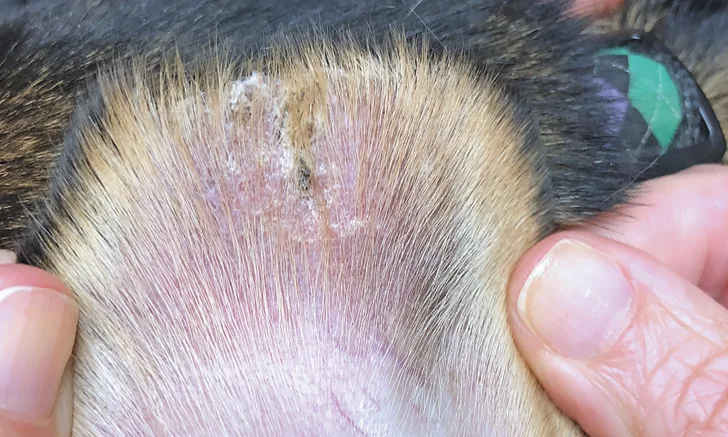
Sebaceous glands typically produce oils and other related substances that keep skin and hair moisturized and lubricated. Skin becomes dry and hair becomes brittle when these glands are missing. Secondary bacterial infection can occur. Lesions can occur anywhere and are typically widespread; however, many affected dogs may initially have lesions only on the pinnal margins, and there have been cases limited to the pinnae.13 Clinical signs include scaly alopecia with hair casting (ie, keratin debris adherent to hairs; Figure 7). In cats, lesions typically start on the pinnae or face.

Sebaceous adenitis causing scaly alopecia on the pinna
Diagnosis of sebaceous adenitis is typically made through skin biopsy and histopathology. Treatment includes topical and systemic therapies. Frequent bathing of the affected dog or cat with moisturizing, keratolytic shampoos is typically necessary. In more severe cases, soaking the dog or cat in diluted propylene glycol or diluted baby oil may be helpful and should be followed by bathing with keratolytic shampoos. Use of a topical blend of essential fatty acids on a weekly basis may also be effective. Cyclosporine is the only systemic treatment of choice shown to result in an increase in sebaceous glands along with clinical improvement.15,16 Other treatment options include omega-3 and -6 fatty acids, vitamin A, and doxycycline or niacinamide.12
Conclusion
Numerous diseases affect the pinnae, including infectious, ectoparasitic, immune-mediated, and neoplastic diseases and keratinization disorders. The clinical conditions discussed here are not an exhaustive list but are common causes of changes to the pinnae.