Top 3 Tips for Intubating Brachycephalic Dog Breeds
Administering anesthesia to dogs considered brachycephalic breeds requires special consideration to accommodate brachycephalic syndrome—the typical anatomic abnormalities that cause some degree of upper airway obstruction (eg, stenotic nares, elongated soft palate, laryngeal collapse, hypoplastic trachea, laryngeal saccule eversion) in these breeds.1
The most common abnormalities seen in brachycephalic breeds are:
Stenotic nares: A condition where the nostrils are too narrow and sometimes collapse inward during inhalation, making it difficult for the patient to breathe through its nose
Hypoplastic trachea: A condition where the diameter of the trachea (ie, windpipe) is smaller than normal
Elongated soft palate: A condition where the soft palate is too long and its tip protrudes into the airway, interfering with inspiration.
Brachycephalic dogs tend to compensate for these respiratory insufficiencies, but sedation and anesthesia remove the compensatory mechanisms2 and it becomes the anesthetist’s responsibility to monitor and protect the patient’s airway.
The following tips will help ensure a smooth experience when preparing a brachycephalic dog for anesthesia.
1. Preoxygenate During Induction
Sedation from premedications can cause brachycephalic patients to experience partial airway obstruction and desaturate quickly. Preoxygenation should occur prior to and during induction. Often the patient benefits from preoxygenation during IVC placement and while attaching monitoring equipment, so long as preoxygenation does not contribute to patient stress. During administration of induction drugs, provide oxygen flow via facemask or nasal oxygen prongs. Many anesthetic induction agents (eg, propofol, alfaxalone) can cause apnea if administered too quickly. The key is to administer the agent slowly (ie, longer than 60 seconds) and only to effect. When possible, follow this sequence of events:
Premedicate/sedate
Preoxygenate
Administer induction medication intravenously
Intubate when the patient is adequately anesthetized.
Administering 100% oxygen before anesthesia induction prolongs the time to onset of arterial hypoxemia3 and increases the body’s oxygen stores, primarily in the functional residual capacity of the lungs.3 Do not preoxygenate if the patient is overly stressed.
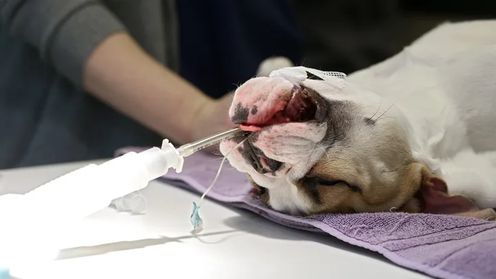
2. Use the Laryngoscope Effectively
The lighted laryngoscope allows the anesthetist to properly visualize the tracheal opening, soft palate, and other oral cavity structures, and to note any abnormalities, such as polyps, masses, or dental abnormalities.
Two types of laryngoscope blades, the Macintosh and the Miller, are commonly used in veterinary medicine.
The Macintosh has a curved blade and is designed to slide into the vallecula (ie, the groove between the base of the tongue and the epiglottis) to move the epiglottis. Because of brachycephalic breeds’ short muzzles, the Macintosh’s blade can help better visualize the laryngeal folds that often are hidden behind the elongated soft palate.
The Miller has a straight blade designed to touch and directly move the epiglottis.4
3. Be Prepared with at Least 3 Sizes of Endotracheal (ET) Tubes
Brachycephalic breeds often require a smaller diameter of endotracheal tube than compared to dogs of similar weight classes. This is often due to the anatomical abnormalities previously mentioned (eg, elongated soft palate, everted laryngeal saccules).
Because of these airway abnormalities, you may have to extubate and reintubate the patient a number of times during the procedure and should always use a clean tube. Therefore, a variety of endotracheal tubes should be available before anesthetizing a brachycephalic patient.
Brachycephalic dogs, because of their hypoplastic trachea and everted laryngeal saccules, often require an ET tube much smaller in diameter than patients of other breeds of similar weight. Clear PVC tubes are preferred over orange rubber ET tubes to better visualize air moving within the tube.3 Clear tubes are also high-volume/low-pressure, which means a larger volume of air in the cuff of the endotracheal tube can be distributed to exert lower pressure on the trachea compared with a similar sized orange rubber ET tube. Red rubber tubes are considered high pressure/low volume and carry the risk of higher pressure within the cuff causing trauma to or necrosis of the tracheal wall.
Conclusion
Brachycephalic breeds’ anatomic differences require the anesthetist to carefully monitor breathing and any airway disturbances. However, proper premedication, vigilant monitoring during the preoperative to recovery stages, and a stress-free induction and recovery can help make these patients less challenging and more rewarding.