Tips & Techniques for Pelvic Radiography
Laura J. Armbrust, DVM, DACVR, Kansas State University
Pelvic radiographs in dogs are commonly obtained to assess a variety of disorders, most commonly hip dysplasia and fractures. The ability to obtain high-quality, correctly positioned radiographs is important for accurate radiographic interpretation.
The most commonly used view is the ventrodorsal hip extended view. The lateral view is useful as the orthogonal view for localization of disease (eg, fracture location or coxofemoral luxation). For most views, but particularly the ventrodorsal hip-extended view, sedation is recommended for both normal dogs and dogs with disease.
The following article explains the methods for obtaining correctly positioned pelvic radiographs, and also discusses selected additional views and their uses. Even though the pelvis is the area of interest, most views generally include the stifles; thus, the x-ray beam is usually centered on the coxofemoral joints or ischium.
Step-by-Step: Pelvic Radiography Positioning
Step 1
Figure 1A demonstrates the normal positioning for the ventrodorsal hip-extended view (the view that should be submitted to the Orthopedic Foundation for Animals). This view is best obtained with the dog placed in dorsal recumbency, which is done by using a foam positioning trough. All but the pelvis and hindlimbs are within the trough. While in a flexed position, the limbs are internally rotated and abducted so that the stifles are almost touching. The limbs are then extended, maintaining the internal rotation, until the femurs are parallel with the table.
If the stifles are kept internally rotated, the patella should be centrally located over the distal femurs, as seen in the normally positioned radiograph (Figure 1B). In this image the femurs are parallel with each other and parallel with the imaging plate. Note the uniform and equal size of the obturator foramen on this well-positioned radiograph. Ideally, the sacrum, ilial wings, and entire 7th lumbar vertebra should be included in the image. Figure 1C shows an example of the pelvis correctly positioned.
Procedure Pearl
Altering the degree of limb extension or flexion and changing the degree of internal or external rotation can significantly affect the appearance of the femoral head and neck. This can be useful when radiographic changes are equivocal. Ensuring that both limbs are positioned similarly allows side-to-side comparisons.

FIGURE 1A
Step 2
Pelvic radiographs must be assessed for adequate positioning and should be repeated if the pelvis is oblique, as in Figure 2A. In this example, there are multiple positioning errors. The right femur/stifle should be further internally rotated (so the patella is more centrally located over the femur). The right stifle should be moved axially (medially) so the femurs are parallel. The pelvis is rotated with the right hemipelvis farther away from the imaging plate. In Figure 2B, the right hemipelvis has been elevated from the imaging plate. In this image, the right (up) obturator foramen is increased in width compared with the left. Additionally, the left (down) ilium appears narrower.
Oblique positioning will result in false assessment of dorsal acetabular rim coverage of the femoral head, as shown in Figure 2C. The dorsal acetabular rim (white arrows) appears to provide more coverage of the right femoral head and less coverage on the left side. When the patient is properly positioned, as in Figure 2D, it is evident that coverage of the dorsal acetabular rim (white arrows) is similar on both sides.

FIGURE 2A
Step 3
The lateral radiograph is obtained with the patient in lateral recumbency and the hindlimbs separated (Figure 3A). A foam positioning device is placed between the limbs or under the "up" leg to keep the femur parallel to the table. The forward limb (typically the limb closest to the imaging plate) is labeled. The corresponding normal lateral radiograph is shown in Figure 3B. In Figure 3C, the femoral head (arrow) of the cranially positioned right limb is luxated craniodorsally.

FIGURE 3A
Step 4
Due to the superimposition of the opposite hemipelvis and proximal femurs on the lateral radiograph, it can be useful to obtain oblique radiographs by rotating the pelvis slightly when the dog is in lateral recumbency. This is done by pushing the limb on the side with suspected pathology dorsally (away from the holder) and pulling the opposite limb/pelvis ventrally (toward the holder), as shown in Figure 4A (white arrows). Dorsal acetabular pathology is best seen if the acetabulum in question is positioned dorsally relative to the normal side. In Figure 4B, an indistinct lucent area is noted just caudal to the acetabulum. The oblique radiograph (Figure 4C), with the right hemipelvis dorsal, clearly shows that the fracture (white arrowhead) does not involve the acetabulum.

FIGURE 4A
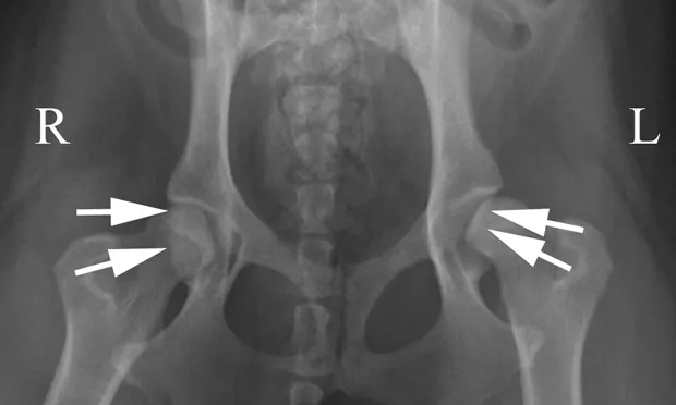
Step 5
The ventrodorsal radiograph with the legs lying in a neutral to flexed position is often called the "frog-legged" position. This view is particularly useful in cases of pelvic trauma because minimal stress is placed on the pelvis and coxofemoral joints. The dog is placed in dorsal recumbency in a foam positioning trough and the limbs are not held. It is still important to maintain symmetry of the pelvis and limbs. Figure 5A demonstrates a dog positioned for this view. Figure 5B shows an example of this positioning in a cat with bilateral capital physeal fractures (white arrows).

FIGURE 5A
Step 6
Distraction techniques can be used to assess laxity of the coxofemoral joints. The PennHIP (University of Pennsylvania Hip Improvement Program) method is the most commonly performed distraction technique, but it requires specialized training. Additional information on this procedure can be found at pennhip.org/PennHIPFAQ.html.
Radiograph submission criteria for the Orthopedic Foundation for Animals can be found at offa.org/hipproc.html.