Microscopic fecal examination is a valuable tool that can help monitor for GI parasitic infections in dogs and cats and should always be included on annual physical examinations.1
Understanding patient lifestyle and possible travel history is key to a fecal examination. Pseudoparasites, spurious parasites, and anomalies should be distinguished and the need for treatment determined. Identifying pseudoparasites and spurious parasites can often be as beneficial for patient well-being as identifying true parasites. To distinguish pseudoparasites and spurious parasites from true parasites observed on initial examination, an additional microscopic fecal examination can be performed after 24 to 48 hours to help confirm the presence of the unknown structure if the patient is not exposed to any source of infection from coprophagy, ingestion of various small animals, or environmental contaminants. Additional diagnostic techniques (eg, coproantigen testing, PCR) can also be useful.
Coprophagy is common in dogs and can include feces of other animals (eg, ruminants, rodents, chickens, wildlife), resulting in ingestion of parasite eggs or oocysts that are unable to continue their life cycle, pass through the dog’s GI tract, and appear on microscopic fecal examination.
Diagnostic results typically show no parasitic ova or a common GI parasite that can be treated with parasiticides and lifestyle changes (eg, avoiding ingestion of other animals, feces, or contaminated water). Parasites that are uncommon in dogs in the United States can also be detected but are occasionally misdiagnosed due to morphologic similarities. Uncommon parasitic infections have been increasing, possibly due to an increase in humans traveling with their pets.
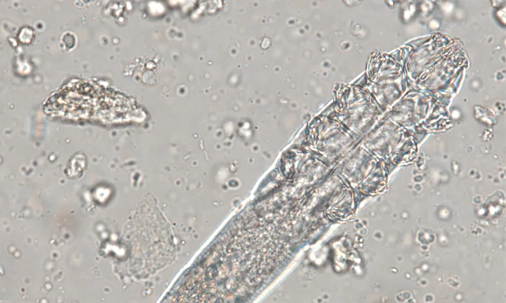
The following images show some true feline and canine parasites, as well as pseudoparasites and spurious parasites that can make diagnosis challenging.

FIGURE 1
Cystoisospora spp oocysts in canine feces are true parasites of dogs. Oocysts in 2 different developmental stages that can help confirm Cystoisospora spp infection can be seen. The oocyst (black arrow) with a single sporocyst is nonsporulated when leaving the host and sporulates over time before becoming infective. A single sporocyst becomes 2 sporocysts (white arrow), each of which contains 4 sporozoites.2