Surgeon's Corner: Tail Fold Caudectomy
J. Brad Case, DVM, MS, DACVS, University of Florida
W. Alex Fox-Alvarez, DVM, MS, DACVS-SA, Community Care Veterinary Specialists, Gainesville, Florida
Presentation
A 4-year-old castrated English bulldog was presented with a 2-year history of pain and recurrent pyoderma secondary to coccygeal vertebral malformation (corkscrew tail). The patient was reported to scoot and twirl on his hindquarters in an attempt to alleviate the discomfort. Brown, purulent discharge had been noted intermittently in the folds of the tail. Antibiotic therapy only provided temporary resolution of clinical signs.
Corkscrew tail amputation (caudectomy) is indicated in patients with severe intertriginous disease, chronic infection, or discomfort attributed to abnormal tail morphology.
Surgery
Perioperative antibiotics (ie, cephazolin 22 mg/kg IV q90 minutes) are initiated 30 minutes before surgery. To minimize contamination of the surgical site, an anal purse-string suture is placed using monofilament suture. Routine hair clipping is followed by sterile preparation of skin around the tail base and in the folds. The patient is positioned in sternal recumbency, with the hindlimbs hanging from the surgical table.
A U-shaped skin incision is made dorsal to the tail down to the level where the tail skin begins to fold inward. This U-shaped flap will facilitate wound closure. The skin incision is continued circumferentially around the tail base. Electrosurgery or Metzenbaum scissors are used to dissect the subcutaneous (SC) tissue dorsally, down to the level of C1-C3 (ie, proposed caudectomy site, usually at or between the normal and ventrally directed coccygeal vertebrae). Once this site is identified, the remainder of SC tissue and fat is dissected circumferentially in the same plane. Palpation of the vertebrae throughout the procedure is useful to guide dissection. Extra caution is taken when dissecting ventrally, as excessive ventral dissection can lead to penetration of the rectum. The dissection plane is maintained ventrally as close to the tail as possible. Once SC tissue is dissected 360<sup°sup>, the area of proposed amputation can be palpated and the intervertebral space identified by manipulation of the tail. Ideally, the cut is made in the intervertebral space; however, mid-body dissection is also an appropriate option. In this case, Ruskin-Liston straight bone cutting forceps were used to perform the amputation.
Major vessels of the tail include the lateral caudal arteries and the median caudal artery, which lies ventral to the tail. In most cases, these vessels can be identified and ligated or cauterized prior to making the cut; however, because the abnormal anatomy of the corkscrew tail can make this difficult, often vessels are cut along with the vertebrae and bleeding is controlled afterward. Small vessel hemorrhage can be managed using electrosurgery, but larger (>2 mm) hemorrhaging vessels should be ligated using absorbable suture. After amputation is complete, the surgical site is first palpated to ensure a smooth cut and then copiously lavaged.
Closure begins with a deep SC layer covering the surgical site, using fine absorbable monofilament suture (ie, poliglecaprone 25) in a buried cruciate pattern. In many dogs, a second SC closure is completed using the same technique. The subcuticular layer is then closed using fine absorbable monofilament suture in a continuous pattern. Finally, the skin is closed using either an intradermal pattern or interrupted external skin sutures. Accurate appositional closure is paramount, as the surgical site is in close proximity to the anus, making it a high-risk region for infection.
The incision can be covered with antibiotic ointment to provide a hydrophobic barrier following skin closure. The patient should wear an Elizabethan collar for 2 weeks (ie, while the incision heals).

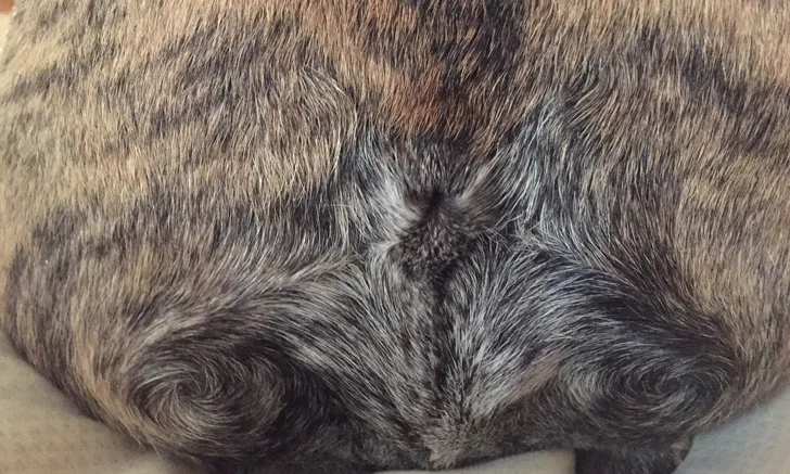
Figure A shows the tail fold caudectomy site 6 months postoperatively.
Outcome
This patient recovered uneventfully and healed well. Only mild erythema of the skin incision was noted on the first 3 postoperative days. The owner considered the outcome to be excellent, as no complications occurred and the dog’s chronic source of pain was removed. Potential complications include infection, dehiscence, delayed healing, and decreased anal tone/sensation. Corkscrew tail amputation has been associated with a good to excellent outcome and low complication rate.1