Staging Melanoma of the Oral Cavity or Skin in Dogs
Diagnosing diseases and conditions may require a various array of diagnostic tests and modalities. This column discusses when a test or procedure is indicated as well as its advantages, disadvantages, economic impact, and reliability of results.Melanomas are typically pigmented tumors of the skin and mucous membranes. Some melanoma tumors may be nonpigmented (amelanotic).
Early studies found that the World Health Organization (WHO) staging system for melanoma (see Box) provided prognostic information,1 but more recent information suggests that these criteria should be combined with information from histopathology (especially the mitotic index).
Current recommendations for staging melanoma include:
Detailed information on the location of the primary tumor
Accurate tumor measurements and lymph node evaluation by cytology or biopsy
Careful evaluation for metastasis by thoracic radiography, CT, or MRI
Evaluation of the histopathology criteria listed above.
While such information will not provide an accurate prognosis for every canine patient with melanoma, it will allow the owner and oncologist to make a more individual treatment decision.
Physical Examination
IndicationsThe physical examination is critical in staging the tumor and establishing some prognostic criteria. One of the most important prognostic factors gained from the physical examination is tumor location.
A recent study distinguished between the behavior of melanomas of the feet and lips and the behavior of those of the skin and oral cavity, respectively. Tumors in haired-skin areas are generally benign (although there are exceptions), while tumors at mucocutaneous junctions, nail bed, and mouth are most often malignant, with the vast majority of oral melanomas being malignant. Median survival and mortality due to melanoma were 22 months and 30%, respectively, among dogs with melanoma of the feet or lips, 5 months and 68% among dogs with oral melanoma, and 24 months and 7% among dogs with cutaneous lesions. When the tumor recurred or metastasized, it did so more rapidly for dogs with oral lesions (median, 2.5 months) than for those with toe/lip melanoma (5.3 months) or cutaneous melanoma (8.9 months).2
Another important prognostic factor gained from the physical examination is the size of the primary tumor. The size of an oral melanoma was associated with survival in a study that used radiation therapy as the primary treatment (Table 1).3
AdvantagesEasy to perform and minimally invasive; provides critical information for staging and prognosis
DisadvantagesNone
Economic ImpactMinimal; it needs to be done for all patients.
Reliability of ResultsPhysical examination findings will often need to be illuminated by further staging (eg, imaging, biopsy) but are reliable if done carefully.
Minimum Database
IndicationsA complete blood count, baseline serum biochemistry panel, and urinalysis (with culture, if indicated) are always recommended for baseline staging of cancer, particularly if surgery, chemotherapy, or multiple anesthesias (such as for radiation therapy) are anticipated.
AdvantagesEasy to perform and minimally invasive; provides critical information for planning treatment and anesthesia
DisadvantagesNone
Economic ImpactMinimal; it needs to be done for all older patients.
Reliability of ResultsResults are reliable if testing is performed on serviced equipment and by qualified personnel.
Lymph Node Assessment
IndicationsThe metastatic rate is very high for oral melanoma, but the time to metastasis varies. At diagnosis, the mandibular lymph nodes (ipsilateral and contralateral) should be palpated and fine-needle aspiration performed for cytologic examination. Since the oral cavity drains to the parotid and retropharyngeal lymph nodes, these nodes can be assessed by CT (and possibly ultrasonography) if advanced imaging is planned.

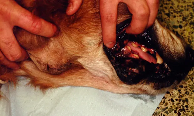
Oral melanomas have a high metastatic rate, as seen in this 14-year-old golden retriever. To accurately assess the primary lesion (white arrows), physical examination may need to be complemented with radiography and, possibly, CT if aggressive resection is planned. Note the enlarged mandibular lymph node (yellow arrows) that should be aspirated for cytology or biopsied along with the primary lesion.
Evidence of atypical melanocytes or melanophages on cytology suggests metastasis. Suspicious results on aspiration cytology should be confirmed by surgical biopsy. Note that although 60% of enlarged lymph nodes had cytologic evidence of metastasis in a study of 100 dogs with oral melanoma, metastases were detected in 40% of normal-size lymph nodes.4Thus, even normal-size lymph nodes should be aspirated or biopsied. The mandibular nodes are the most commonly affected site for oral melanoma; however, when melanomas of other sites are diagnosed, the regional lymph nodes should be similarly assessed.
AdvantagesEasy to perform and minimally invasive; provides critical information for staging and prognosis
DisadvantagesCytology results may be equivocal and biopsy may be required to definitively determine whether metastasis is present.
Economic ImpactCost of cytology and procedure
Reliability of ResultsCytology is reliable if fine-needle aspiration is performed carefully, but "negative" results could be misleading.
Biopsy
IndicationsAlthough histopathology is required for definitive diagnosis of melanoma, the index of suspicion should be high in an old dog with a friable oral mass or a dog with a cutaneous (especially digital) pigmented lesion. For most melanomas, the diagnosis may be made by finding melanin granules in the cytoplasm on light microscopy. Some immunohistochemical markers (S-100, Melan-A, HMB-45) have been explored for their value in diagnosing melanoma when the tumor is poorly pigmented and poorly differentiated.
A recent study found shorter survival for dogs with melanomas from any site that was larger, had high numbers of mitotic figures per 10 high-powered fields, had marked nuclear atypia, and had more inflammation or necrosis (these characteristics were used to calculate a numeric score; see Table 2).2
Biopsy of the regional lymph nodes, even those not enlarged, at the time of lesion biopsy is recommended because it will affect staging and may direct therapy. One study found that dissection of the lymph nodes at surgery increased the number of dogs diagnosed with metastatic disease.5
AdvantagesCan be performed safely and provides critical information for staging and prognosis
DisadvantagesRequires general anesthesia and careful planning so as not to negatively affect future definitive surgery attempts; best to consult with the surgeon or radiation oncologist who will be performing definitive treatment
Economic ImpactCosts of anesthesia, procedure, and pathology, as well as potential additional costs of immunohistochemistry if needed
Reliability of ResultsExtremely reliable when sample is properly collected and fixed
Imaging of Tumor & Distant Sites
IndicationsFine-detail radiographs of the affected area, including the digit or the dental arcade (depending on site of primary tumor), provide information on the size and invasiveness of the melanoma. Radiographs of the affected digit show lysis of P3 in approximately 80% of dogs with subungual squamous cell carcinoma. This is in contrast to digital melanomas, which rarely cause bony destruction (5% of cases). CT or MRI will more accurately delineate bone involvement and should be considered if the tumor margins are uncertain on physical examination or radiography.
Thoracic radiographs may indicate pulmonary metastasis at the time of diagnosis and should be obtained before definitive treatment. Pulmonary metastasis, however, frequently occurs late in the disease.6,7 Thoracic CT is more sensitive at detecting pulmonary metastases and should be considered when access is possible and finances permit.Melanoma may also spread systemically to other sites, and metastasis has been reported in the kidney, myocardium, and brain.8 If involvement of other sites is suspected (based on results from baseline blood analysis or physical examination and history), then imaging (abdominal ultrasonography, CT, or MRI) of the abdomen or central nervous system is warranted.
AdvantagesCan be performed safely and provides critical information for staging and prognosis
DisadvantagesMay require sedation for radiography and ultrasonography; will require general anesthesia for CT or MRI, as well as access to facilities. Radiography and CT carry a risk for radiation exposure. It is worth consulting with the surgeon or radiation oncologist who will be performing the definitive treatment to ensure correct views are obtained.
Economic ImpactCosts of anesthesia and procedure can be considerable, but these tests will allow for more accurate treatment decisions, minimizing risks for toxicity or inadequate treatment of tumor.
Reliability of ResultsExtremely reliable if performed on serviced equipment and by qualified personnel.