In the Literature
Rich N, Brune J, Duclos D. A novel cytological technique for bacterial detection on canine skin. Vet Dermatol. 2022;33(2):108-e30. doi:10.1111/vde.13036
The Research ...
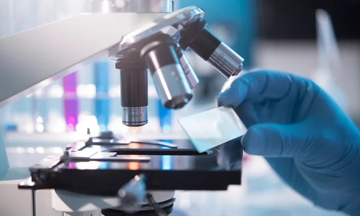
Impression smear and tape-strip preparations are traditional and validated skin cytology methods for diagnosis and monitoring of suspected bacterial or Malassezia spp overgrowth.
This study compared slurry preparation, a novel cytologic sampling method, with traditional methods to detect bacteria and Malassezia spp in 30 dogs presented with atopic dermatitis that had lesions consistent with superficial bacterial pyoderma and/or Malassezia spp dermatitis. Samples were collected using impression smear, tape-strip, and slurry preparation methods and stained with modified Wright-Giemsa stains.
For slurry preparation, a microspatula with a flat-ended blade was used to scrape the surface of a lesion; debris (including scale and crust) was collected on a glass slide. One drop of sterile water was placed on the slide and gently mixed via rocking. The slide was then briefly heated on a hot plate, after which the slurry was mixed and larger debris macerated with a wooden applicator stick, yielding an opaque liquid. Larger, unmacerated debris was removed from the sample, and the preparation was again briefly heated to dry remaining water. This preparation method took 2 to 3 minutes.
Slurry preparation identified significantly higher numbers of bacteria compared with other techniques; however, tape-strip cytology detected more yeast than slurry preparation.
Slurry preparation is a reasonable alternative to impression smears and tape-strip preparation for crusted and scaly lesions to improve chances of identifying bacterial infection. The authors recommend sampling pustules with impression smears instead of the slurry method, as pustules need to be ruptured prior to sampling.
... The Takeaways
Key pearls to put into practice:
Skin cytology is recommended at both initial and follow-up examinations in patients presented for itching, scaling, crusts, or skin debris.1,2 A combination of sampling methods can be used, depending on lesion appearance. For example, tape-strip cytology may be used on inflamed and scaly feet, impression smears may be used for a pustule on the ventral abdomen, and slurry preparation may be used for crusting on the dorsum during a single examination of a dog.
It may be easier for less experienced examiners to review impression smears and slurry preparations for bacteria, Malassezia spp, and inflammatory cells; tape-strip cytology preparations can appear more crowded to the untrained eye.3
Skin cytology allows for judicious oral antimicrobial use, as patients may have scaling or crust caused by Malassezia spp infection alone.3 Routine use of skin cytology at follow-up examinations can also help guide timing of bacterial culture and enable correlation between culture results and morphologic characteristics of bacteria on cytology.1
You are reading 2-Minute Takeaways, a research summary resource presented by Clinician’s Brief. Clinician’s Brief does not conduct primary research.