Skin Clues to Canine Endocrine Disease
Rosanna Marsella, DVM, DACVD, University of Florida
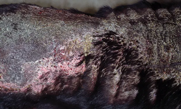
Figure 1 Dog with iatrogenic calcinosis cutis. Note the thick plaques on the dorsal neck and trunk. Calcinosis cutis starts with a papular eruption that can progress to thick plaques of calcification and mineralization.
In dogs, endocrine disease can manifest with a variety of cutaneous signs. In some cases, signs may not be disease specific; for example, both Cushing’s disease and hypothyroidism can trigger demodicosis, secondary skin infections (bacteria and yeast), and truncal alopecia with comedones (blackheads). In comparison, certain signs can be identified with a disease process, such as rat tail in dogs with hypothyroidism; in these patients, alopecia is primary and not secondary to pruritus, as is observed in dogs with flea allergy.

Figure 2 Yorkshire terrier with alopecia X. Note the truncal hyperpigmentation and alopecia. This disease typically spares the head and extremities.
Calcinosis cutis (Figure 1), a peculiar cutaneous manifestation of Cushing’s disease seen only in dogs, presents as an intensely pruritic papular dermatitis typically affecting the dorsal neck and trunk and occasionally affecting frictional (eg, axillary, inguinal) areas. The papules are hard on palpation and have a white central area. By contrast, different manifestations are presented in other species affected by Cushing’s disease. For example, cats may develop a syndrome of extreme skin fragility in which skin can tear easily despite minimal trauma.In dogs with a condition referred to as alopecia X or pseudo-Cushing’s (Figure 2), truncal alopecia and hyperpigmentation are manifested without the other systemic signs seen with Cushing’s disease; that is, alopecia is progressive but spares the head and extremities. Alopecia X is believed to be linked to sex hormone imbalances and, in most cases, is considered a cosmetic condition that does not require treatment. While the cause is still under investigation and may be multifactorial, alopecia X is likely the result of an imbalance of adrenocortical steroid hormone intermediates.1
Both calcinosis cutis and alopecia X are examples of how the skin can be an important window to internal organs, providing key clues in the diagnosis of systemic endocrine disease.