Seizures, Lethargy, & Collapse in a Dog
Suzi, a 9-year-old spayed female hound mix, was referred for a 6-month history of seizures that were unresponsive to treatment with phenobarbital.
History
Suzi’s owners also reported that she had frequent episodes of lethargy and collapse. More recently, they noticed she was urinating frequently and eating and drinking more than usual, and were concerned that she might have diabetes.
Physical Examination
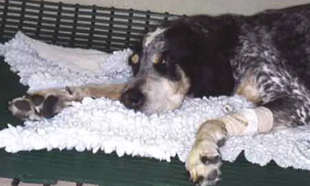
Suzi was quiet, alert, and responsive (Figure 1/See photo at the top of the page). Body condition score was 6/9. A complete neurologic examination showed no abnormalities, but a facial twitch developed during the examination. Cranial abdominal organomegaly was noted. The remainder of the physical examination was unremarkable. The facial twitching resolved after she was fed baby food containing chicken.
Laboratory Results
Results of a complete blood count and urinalysis were unremarkable. Serum biochemical profile results (Table 1) showed hypoglycemia and increased concentrations of alkaline phosphatase; other results were within normal limits. Serum glucose and insulin concentrations are listed in Table 2.
Figure 2: Ultrasound image of the pancreatic mass; the pancreatic mass is a hypoechoic irregular nodule outlined by the + and x markers

Diagnostic Imaging
Thoracic radiographs showed no abnormalities. Abdominal ultrasound revealed a hyperechoic nodule in the liver, and the liver had rounded margins (image not shown). The pancreas had enlarged irregular borders with mixed echotexture and a mass in the right lobe (Figure 2).
Ask Yourself...
Assuming that the data are most consistent with insulinoma, what is the best course of action?
A. Conservative management (frequent small meals and prednisolone) because surgery does not improve survival timeB. Exploratory laparotomy to remove the pancreatic mass and liver noduleC. Weekly injections of long-acting corticosteroids and frequent evaluation of insulin concentrationsD. Treatment with exogenous insulin to suppress release of endogenous insulinE. Treatment with corticosteroids and diazoxide
Correct answer:
B. Exploratory laparotomy to remove the pancreatic mass and liver nodule
Insulinomas are insulin-secreting tumors of the pancreatic beta-islet cells. Diagnosis is suggested by a history of lethargy and collapse or seizures, often associated with exercise.
Diagnosis
Observation of hypoglycemia with simultaneous hyperinsulinemia is highly suggestive of an insulin-secreting tumor. The presumptive diagnosis is also often made by observation of low fasting blood glucose in the presence of normal concentrations of insulin in the peripheral blood; however, definitive diagnosis is only obtained via histopathology.1
Other tumors that may cause hypoglycemia include leiomyomas, leiomyosarcomas, and hepatocellular tumors.2 Abdominal ultrasound may be helpful in detecting tumors of non–beta-cell origin, although an inability to detect a pancreatic mass on ultrasound does not preclude the diagnosis of an insulinoma.1,2 Similarily, detection of a pancreatic mass does not confirm the presence of neoplastic disease.1,2
Treatment
Approximately 50% of dogs have metastasis at the time of diagnosis, typically involving the liver and regional lymph nodes.2 Surgery to remove the pancreatic mass results in significantly improved survival times when compared with conservative management. A recent study documented a median survival time of 547 days in dogs treated conservatively versus 785 days in dogs treated with surgery and 1,316 days in dogs that had surgery followed by medical treatment of relapses.3
Surgical Management
Surgical management involves exploratory laparotomy with partial pancreatectomy. Approximately 14% of these tumors are found in the body of the pancreas with an even distribution throughout both limbs.4 Tumors in the body of the pancreas may prove difficult to remove without disrupting the duodenal or pancreatic blood supply. Marginal excision of insulinomas is satisfactory, and removal of a large amount of adjacent normal tissue is not necessary.
Removal of pancreatic masses can be achieved either by careful dissection and ligation of the blood vessels supplying the tumor, or by placement of multiple sutures around the tumor to crush the parenchyma of the pancreas while ligating vessels and small pancreatic ducts. Regional lymph nodes and any gross metastases seen at exploratory laparotomy should be removed if possible.
Figure 3: Intraoperative appearance of the pancreas and pancreatic mass: the mass is the darkened area indicated with the black arrow; the normal pancreatic tissue is indicated by the asterisk.

The mass found in Suzi is pictured in Figure 3. Even small masses that may be difficult to appreciate grossly can secrete significant amounts of insulin. Surgical removal of the primary tumor and accessible metastatic foci increases survival time even in the presence of gross metastatic disease.1
Medical Management: Insulinoma or Postoperative Recurrence of Hypoglycemia
Small, frequent meals high in protein, fat, and complex carbohydrates
Limited exercise
Prednisolone (0.25mg/kg Q 12 H), titrated to effect dependent on blood glucose levels
± Diazoxide (5mg/kg Q 12 H), introduced gradually; monitor patient response
Postoperative Care
Ten percent to 43% of surgically managed cases will have pancreatitis after surgery.2 Animals are typically preemptively managed for postoperative pancreatitis by withholding food for 24 hours after surgery and administering intravenous fluids and antiemetics.2 Parenteral nutritional support may be necessary in some extreme cases.
Animals may have persistent hypoglycemia postoperatively or develop rebound hyperglycemia, and careful monitoring of glucose concentrations is recommended.2
Figure 4A: Intraoperative appearance of the liver nodule, marked by the black arrow

Figure 4B: Cut section of the liver nodule showing the metastatic lesion (B)

Prognosis
Reports in the literature vary; however, clinical signs typically recur in 50% of dogs within 18 months after surgery.2 Prognosis for regression of clinical signs is higher for dogs treated surgically than for those receiving conservative treatment in all reports.1-4
Outcome
he pancreatic mass found in Suzi was removed along with the liver nodule via a partial liver lobectomy (Figure 4). Histopathologic evaluation confirmed a beta-islet cell tumor with metastasis to the liver. She became euglycemic after surgery and was discharged without any incident.