Respiratory Distress & Intermittent Coughing in an Italian Greyhound
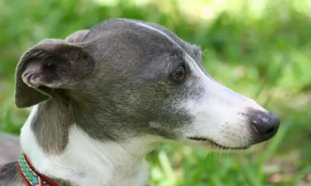
An 11-year-old, spayed female Italian greyhound presented for evaluation of a 24-hour history of increased respiratory rate and effort combined with intermittent coughing.
History. The dog was evaluated for decreased appetite and weakness 1 ½ weeks earlier, at which time an increased rectal temperature of 104° F (40° C) was noted. Enrofloxacin treatment was initiated.
When clinical signs did not improve after 5 days, the enrofloxacin was replaced with amoxicillin-clavulanic acid and deracoxib. The decreased appetite progressed to anorexia, coughing was noticed by the owner at night, and respiratory rate and effort worsened.
Physical Examination. The patient was quiet, alert, and responsive, with a respiratory rate of 80 breaths/min. Her increased respiratory effort improved with supplemental oxygen. Mucous membranes were light pink, injected, and tacky with a capillary refill time of 1 second.
Diffusely harsh lung sounds, pulmonary parenchymal crackles in the right ventral thorax, grade 3/6 left-sided apical systolic heart murmur, tachycardia (heart rate, 180 bpm), and weak synchronous dorsal metatarsal pulses were evident. The rectal temperature was decreased at 97.4° F (36.33° C). The dog was approximately 7% to 8% dehydrated.
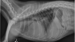
Diagnostics. The patient’s Doppler blood pressure was 40 mm Hg systolic, and pulse oximetry was 94% on 40% FiO2. Significant blood analysis findings are summarized in the Table and thoracic radiographs taken at presentation are shown in Figure 1.
Figure 1: Thoracic radiographs: Right lateral (A), left lateral (B), and dorsoventral (C)

_FiO2 = fraction of inspired oxygen; PaO2 = arterial oxygen partial pressure; PaCO2 = arterial carbon dioxide partial pressure
_

Ask Yourself...
What are the most likely differential diagnoses based on the findings?
What major changes are evident on thoraic radiography?
What therapeutics should be initiated during hospitalization?
What continued care might be needed upon discharge?
DIAGNOSIS: Pneumonia, probably bacterial in origin
Radiography and complete blood count (CBC) findings were consistent with a diagnosis of pneumonia, most likely bacterial in origin. The dog’s history was not typical of aspiration pneumonia, as no prior vomiting episodes were noted by the owner.
CBC = complete blood count; FiO2 = fraction of inspired oxygen
Based on the heart rate, weak pulses, lactate findings, and blood pressure, the dog was in shock, probably from a combination of hypovolemia, sepsis, and hypoxemia. The dehydration was likely secondary to anorexia but may have been related to free water loss through increased respiratory rate and effort, which was supported by a finding of increased serum sodium.
Stabilization. Because the patient was cardiovascularly unstable on presentation and had a significant heart murmur, initial treatment involved 2 conservative IV crystalloid fluid boluses (10 mL/kg each) for hypovolemic shock, oxygen supplementation, and ongoing crystalloid fluid administration for dehydration. Respiratory rate and effort improved in an oxygen cage with the FiO2 set at 50%, and heart rate and blood pressure normalized with fluid therapy.
Additional Diagnostics. With the dog’s history of prior broad-spectrum antibiotic therapy, obtaining a sample from the lungs for bacterial culture and sensitivity testing was imperative. Because of respiratory and cardiovascular instability, however, placing the dog under general anesthesia for an endotracheal wash was considered too dangerous, and the patient’s small size precluded a transtracheal wash.
Since consolidation of the lung was present on radiographs and only a small amount of pleural effusion was present, ultrasound-guided needle aspiration of the lung was performed under local anesthesia to obtain a small sample of pleural fluid. The dog received 10 mg/kg clinda-mycin IV Q 12 H and 40 mg/kg cefotaxime IV Q 6 H while culture and sensitivity were pending.
Cytology of the pleural fluid and the lung aspirate yielded identical results showing chronic severe suppurative inflammation. Culture yielded a multiple–drug-resistant Enterococcus faecium that was sensitive to chloramphenicol.
Did You Answer...
Pneumonia and congestive heart failure are the most likely diagnostic differentials.
Given the dog’s respiratory distress, tachycardia, heart murmur, hypotension, hypoxemia, and decreased rectal temperature, left-sided congestive heart failure would be a differential diagnosis.
However, the dog’s left-shifted CBC with toxic change and the distribution of the alveolar pattern on thoracic radiographs make this diagnosis less likely.
The presence of pleural effusion, which is atypical for bronchopneumonia in dogs, combined with the severity of alveolar consolidation, would also make a necrotic pulmonary mass or abscessed lung lobe with subsequent rupture into the pleural space other possible differentials.
Alveolar pulmonary pattern associated with (1) air bronchograms and (2) involving the right cranial, right middle, and left cranial lung consistent with extensive bronchopneumonia are the major radiographic findings. Based on the distribution, the pattern is most likely secondary to aspiration.
Treatment for compromised patients with bronchopneumonia includes:
Oxygen therapy and intravenous fluid therapy (crystalloids and/or colloids) to maintain hydration and keep the respiratory secretions moist.
Intravenous antibiotics are advised in hypoxic or hypovolemic shock patients because perfusion of the gastrointestinal tract, muscle, and subcutaneous tissues can be variable and cause unpredictable absorption.
Other in-hospital therapeutics may include nebulization with sterile saline, gentle coupage, a mucolytic agent such as N-acetylcysteine, and bronchodilators.
For this dog, intravenous crystalloid and colloid therapy, oxygen, nebulization and coupage, intravenous chloramphenicol, intravenous N-acetylcysteine, and intravenous aminophylline were the elected treatments.
Home care should involved continued antibiotic therapy, strict rest, restricted access to other dogs, and continued nebulization and coupage.
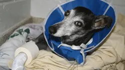
Treatment. Once the patient was stabilized, treatment involved ongoing oxygen and fluid therapy, intravenous antibiotics, as well as nebulization (Figure 2) and coupage. In compromised patients, antibiotic therapy should be based on culture and sensitivity testing (see TX at a Glance). In addition, administration of mucolytic agents can help reduce the viscosity of airway secretions.
Figure 2: The patient receiving nebulization; a 2-inch roll of tape is placed at the end of the nebulization tubing to stabilize it
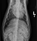
Before the patient was released to home care, follow-up thoracic radiographs (Figure 3) were obtained and compared with the radiographs taken on presentation to confirm sufficient improvement for hospital discharge.

Figure 3: Thoracic radiographs taken before discharge from the hospital show improvement, but not complete resolution, of alveolar disease:

Right lateral (A), left lateral (B), and ventrodorsal (C)
Home Care. In addition to oral chloramphenicol, other treatment recommendations for continued recovery at home included strict rest, restricted access to other dogs, airway humidification at home for 10 minutes 2 to 3 times daily
(accomplished by enclosing the dog in a bathroom and running a hot shower to saturate the inspired air with water vapor), along with gentle coupage 4 to 6 times per day.
Outcome. The dog returned every 2 weeks for follow-up radiography, and radiographic resolution of the pneumonia was evident at 4 weeks. Chloramphenicol therapy was continued for an additional 2 weeks beyond radiographic resolution of pneumonia. At recheck 1 month after antibiotic therapy had ended, the dog continued to do well.
TX At A Glance
Supplemental oxygen therapy via nasal oxygen, oxygen cage, or for severely compromised patients, mechanical ventilation
Intravenous fluids to maintain perfusion, hydration, and moist airway secretions
Intravenous antibiotics for compromised patients based on culture and sensitivity testing. Transition to oral antibiotics can be made once cardiovascular stability is achieved and the patient is no longer vomiting or hypoxic and is eating reliably on its own.
Administration of intravenous mucolytic agents, such as N-acetylcysteine, helps reduce the viscosity of airway secretions by reducing disulfide linkages in mucoproteins.
RESPIRATORY DISTRESS & INTERMITTENT COUGHING IN AN ITALIAN GREYHOUND • Dana L. Clarke & Lesley G. King
References
1. Aspiration pneumonia. Barton L. In King LG (ed): Textbook of Respiratory Disease in Dogs and Cats—Philadelphia: WB Saunders, 2004, pp 422-430.2. Bacterial pneumonia in dogs and cats. Brady CA. In King LG (ed): Textbook of Respiratory Disease in Dogs and Cats—Philadelphia: WB Saunders, 2004, pp 412-421.3. Clinical, clinicopathologic, and radiographic findings in dogs with aspiration pneumonia: 88 cases (2004-2006). Kogan DA, Johnson LR, Jandrey KE. JAVMA 233:1742-1747, 2008.
Etiology and clinical outcome in dogs with aspiration pneumonia: 88 cases (2004-2006). Kogan DA, Johnson LR, Sturges BK, et al. JAVMA 233:1748-1755, 2008.
Fine needle aspiration. Cole SG. In King LG (ed): Textbook of Respiratory Disease in Dogs and Cats—Philadelphia: WB Saunders, 2004, pp 135-137.6. Oxygen supplementation and humidification. Tseng LW, Drobatz KJ. In King LG (ed): Textbook of Respiratory Disease in Dogs and Cats—Philadelphia: WB Saunders, 2004, pp 205-213.