Repositioning or Enucleation of the Proptosed Globe
You have asked...
Is the patient with traumatic proptosis of the globe a candidate for repositioning or enucleation?
The expert says...
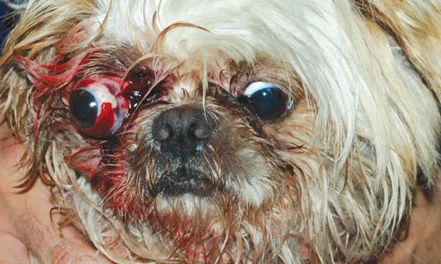
Proptosis of the globe (Figure 1) is a traumatic event for the patient, but it can also be difficult for the client who is likely distressed at the appearance of the eye. Owners of pets with traumatic proptosis of the globe will want to know whether the eye and/or vision can be saved.
The aim of this article is to help the clinician determine whether a patient is a candidate for enucleation or reduction of the globe.
Related Article: Proptosis Reduction
Figure 1. Traumatic globe prolapse in a dog. Note the lateral strabismus, caused by avulsion of the medial rectus muscle. Courtesy of Marta Leiva, Autonomous University of Barcelona

Author Insight
If a client calls and describes globe proptosis in a patient, instruct the client to keep the globe moist using a lubricant or wet gauze while the patient is brought to the closest veterinary care available.
Prognostic Indicators
Several factors can affect the prognosis for vision or for retaining the globe in cases in which vision cannot be salvaged. A review of each of these factors can help the clinician answer the client’s questions and determine the best course of treatment.
Skull Conformation
Traumatic proptosis is common in brachycephalic dog breeds because of the shallow orbit and poor eyelid closure.1,2 In these breeds, minimal trauma may cause proptosis, frequently without additional injuries to the eye, skull, or body, so the overall prognoses for both vision and globe retention are good.
However, in cats and in mesocephalic and dolichocephalic dogs, the eye is situated in a deep orbit and is protected by tight eyelid closure. In these patients, more profound trauma is usually required to cause proptosis, so it is frequently accompanied by other intraocular or bodily injuries. Therefore, prognosis for both vision and globe retention is poorer.3
Duration of Proptosis
As with every medical emergency, prognosis depends on timely medical attention. In proptosis, the eyelids cannot close over the globe; this leads to corneal exposure and desiccation. Depending on duration of the proptosis, patients may be presented with corneal ulceration, necrosis, or perforation, all of which (with the exception of superficial ulceration) will gravely impact the prognosis.
Related Article: Top 5 Emergencies Requiring Anesthesia
Intraocular Injury
Hyphema is a poor prognostic indicator for vision, as it generally only occurs with coinciding trauma to the uvea. The trauma may also cause retinal detachment, lens luxation, or both, but these conditions may be obscured in cases with concurrent hyphema.
Ultrasonography may aid in visualizing such intraocular conditions in cases in which internal structures cannot be seen because of hyphema. Patients with retinal detachment carry a markedly poor visual prognosis. A veterinary ophthalmologist should be consulted in cases of lens luxation.
Globe rupture is another cause of hyphema, and it should also be suspected in cases of collapsed globes, even without apparent injury to the anterior segment. Rupture may be present posteriorly among muscle attachments or adjacent to the optic nerve. Ultrasonography may aid in the diagnosis. The prognosis for salvaging ruptured globes is markedly poor.3-5
Pupils
It is difficult to provide visual prognosis based on the resting pupil diameter (miotic or mydriatic); however, pupillary light reflex (PLR) is an important sign. The presence of a PLR in the proptosed eye is a favorable prognostic indicator for retinal function and the potential for vision. If the pupil cannot be seen, the dazzle reflex in the affected eye and the consensual PLR from the affected eye to the unaffected eye should be assessed.3,6
The presence of a PLR in the proptosed eye is a favorable prognostic indicator for retinal function and the potential for vision.
Strabismus and Extraocular Muscles
Many patients with proptosis are presented with strabismus caused by tearing (avulsion) of extraocular muscles. The medial rectus muscle is usually the first to be avulsed, and affected patients will have a lateral strabismus (Figure 1, above).
Other types of strabismus may indicate avulsion of different extraocular muscles, which is prognostically important. If there is avulsion of 3 or more rectus muscles, blood supply and innervation to the anterior segment of the eye are compromised. This can lead to a grave prognosis for globe salvage. The prognosis for salvaging the globe is guarded when 2 muscles are avulsed, and it is fair if 1 or no muscles are avulsed.2-6
In General
Overall, the prognosis for vision in dogs with traumatic proptosis is guarded, but the prognosis for cosmetic salvage is better. A retrospective study examining 66 cases of proptosis in dogs reported vision in 18 eyes following repositioning and blindness with an intact globe in another 26 eyes. Four patients were euthanized, and 18 eyes were enucleated.7
As noted previously, the prognosis for cats is poorer. In the same study, only 2 of 18 cat eyes were salvaged, and neither remained visual. Twelve of 18 eyes were enucleated, and 4 cats were euthanized.7
Proper counseling of clients about potential outcomes is important. Most clients will be satisfied if the eye can be retained, even if vision in that eye is lost. Therefore, globe reduction should be attempted, and postoperative medical therapy should be directed to preserve the eye unless an extremely poor prognostic indicator (eg, corneal perforation, globe rupture, avulsion of 3 or more extraocular muscles) is present. However, the owner should be warned in advance that a second surgery may be necessary if the reduction fails and enucleation is required. When properly educated and warned, many clients will opt for globe reduction.
Prompt intervention and careful examination to identify prognostic indicators are critical for achieving an optimal outcome for both patient and clients.
RON OFRI, DVM, PhD, DECVO, is associate professor of veterinary ophthalmology at his alma mater, Hebrew University of Jerusalem, where he was a member of the charter class. Winner of numerous Teacher of the Year awards, Dr. Ofri has written more than 60 reviewed papers and is a popular international speaker, contributing author to Veterinary Ophthalmology, and coauthor of Slatter’s Fundamentals of Veterinary Ophthalmology. He received his doctorate from University of Florida, where he studied retinal function in glaucoma.