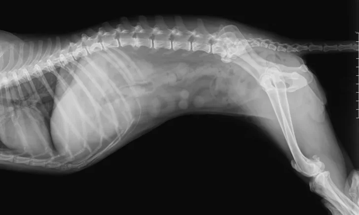
Repeat Radiography: The Mechanical Obstruction Dilemma in Veterinary Patients
James Howard, DVM, MS, DACVS-SA, COVE Emergency Pet Care, Delaware, Ohio

In the Literature
Miles S, Gaschen L, Presley T, Liu CC, Granger LA. Influence of repeat abdominal radiographs on the resolution of mechanical obstruction and gastrointestinal foreign material in dogs and cats. Vet Radiol Ultrasound. 2021;62(3):282-288.
The Research …
Distinguishing mechanical obstruction from functional ileus can be challenging. Radiography is commonly used to evaluate intestinal obstructions, but ultrasonography is used in some clinics and has greater sensitivity than radiography for detection of small intestinal obstructions.1,2 Radiographic features of obstruction include variability in small intestinal diameter, visualization of foreign material, and gas pattern abnormalities.1-4 Clinical signs should be considered in conjunction with subjective appearance of the bowel.5
This retrospective study aimed to determine the incidence of radiographic resolution (ie, absence of foreign material, movement of foreign material into the colon, absence of previously diagnosed intestinal dilation) of obstruction or GI foreign material with medical management. Radiography was repeated within 36 hours of initial radiography, and resolution was 26.8%. A second repeat set of radiography performed 36 hours after initial radiography had resolution of 34.6%, and resolution was 50% following a third repeat set of radiography; however, there was no statistical difference in resolution between the second and third sets.
Foreign material was definitively diagnosed in 46.4% of cases; 29.6% of which were resolved with medical management. A similar rate of resolution (35.5%) was noted in cases with questionable foreign material. When specific dilation patterns were examined, 37.5% of resolved cases were gastric only. Only 17.1% of patients with small intestinal dilation and mechanical ileus had resolution, and only 11.4% of patients with both small intestinal and gastric dilation had resolution. Anatomic location of foreign material was not a significant predictor of resolution.
These results offer a helpful indicator of timing for surgical intervention, as it is illustrated that multiple radiographs 36 hours after an initial study provided no difference in resolution when compared with first repeat radiographs taken within 36 hours of the first study. Lack of radiographic improvements in conjunction with clinical signs and holistic patient evaluation can indicate surgery should be performed earlier, as opposed to pursuing additional radiography.
… The Takeaways
Key pearls to put into practice:
Repeat radiography may be necessary if a mechanical obstruction is not conclusively present: one additional set within 36 hours is recommended. At this author’s institution, repeat radiography within 12 to 18 hours is standard. Surgical exploration is generally recommended when there is radiographic evidence and/or clinical signs of a mechanical obstruction.
Definitive diagnosis of foreign material is made in ≈50% of patients. Clinical signs, patient history, and repeat imaging are paramount to determine surgical candidacy.
Pet owner education is important, as continued diagnostics and medical management incur additional costs. Owners should understand that only ≈30% of patients with clinical signs or suspected mechanical obstruction show radiographic resolution, regardless of whether foreign material is definitively detected.