Regurgitation or Vomiting?
Alex Gallagher, DVM, MS, DACVIM (SAIM), Columbia Veterinary Emergency Trauma and Specialty, Columbia, South Carolina
How do I differentiate regurgitation from vomiting, and what is the best approach for patients with regurgitation?
Vomiting is a common presentation to veterinarians. While the majority of cats and dogs are truly vomiting, some may actually be regurgitating, and owners are often unaware of the difference.
Veterinarians must be able to distinguish these two clinical signs, as the differential diagnosis and evaluation vary considerably for each. The following discusses how to differentiate regurgitation from vomiting and outlines the common causes, diagnosis, and treatment of regurgitation.
Distinguishing the Signs
Regurgitation is the passive expulsion of food, fluid, or other material from the pharynx or esophagus, while vomiting is an active expulsion of ingesta from the stomach and (sometimes) duodenum. In contrast to regurgitation, vomiting involves a centrally mediated reflex with coordinated closure of the nasopharynx and glottis to protect the airway, reducing the risk for aspiration pneumonia. Both of these processes must be differentiated from expectoration (ie, expulsion of material associated with coughing).
A complete history and thorough examination will usually allow differentiation of regurgitation from vomiting (see Table). Asking owners to describe the episodes is often helpful. Abdominal contractions (retching) or bile in the vomitus is specific for vomiting. In addition, these patients may show prodromal signs of nausea (eg, salivation, lip smacking). The clinician should also consider that a patient may be both vomiting and regurgitating, although this is rare.
Clinical Signs That Differentiate Regurgitation from Vomiting
With regurgitation, owners typically report that the patient simply lowers its head and material comes out. Owners may also report finding fluid or food on the floor without hearing the pet vomit. If the owner’s description of episodes does not allow clear-cut distinction, then the rest of the history and examination should determine whether vomiting or regurgitation is more likely.
Timing of the episodes in relation to feeding and the amount of material produced are not distinguishing factors. Patients may vomit undigested food or regurgitate food that appears digested.
The pH of the expulsed material has been considered a possible differentiating test in that vomited material is expected to have a low pH and regurgitated material to have a more neutral or higher pH. However, some patients may regurgitate stomach contents (eg, in reflux esophagitis) and some may vomit bicarbonate-rich fluid refluxed from the duodenum, making pH a poor indicator.
Because regurgitation is uncommon in cats, the diagnosis is more likely to be vomiting. If possible, having the owner videotape an episode may be helpful.
Causes of Regurgitation
Esophageal diseases are the most common cause of regurgitation and can be categorized as obstructions or motility disorders (see Esophageal Conditions Causing Regurgitation, below). Common causes of obstruction include foreign bodies, strictures, or vascular anomalies (eg, persistent right aortic arch). Less common causes include esophageal or paraesophageal tumors.
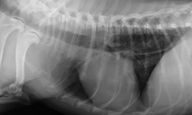
The most common cause of motility disorders is idiopathic disease, either congenital or acquired. Acquired disease may be caused by hypoadrenocorticism, esophagitis, lead toxicity, organophosphate toxicity, or myasthenia gravis (Figure 1). Hypothyroidism may be associated with a motility disorder; however, a definitive relationship has not been established.
Pharyngeal dysphagia, a less common cause of regurgitation, may be suspected based on history, examination, or observation of the pet eating or drinking. Clinical signs include difficult or painful swallowing (which may also be seen with esophagitis), dropping food from the mouth or water from the nostrils, or coughing/gagging when attempting to swallow. Pharyngeal dysphagia may be caused by neuromuscular disorders, pharyngeal tumors, anatomic abnormalities, or trauma (foreign bodies, ingestion of caustic substances).
Esophageal Conditions Causing Regurgitation
Diagnostic Approach to Regurgitation
Plain thoracic and cervical radiography is the initial diagnostic step. Radiographs can help identify megaesophagus (focal or generalized), foreign body, or paraesophageal tumor. In addition, the lungs can be evaluated for signs of aspiration pneumonia.
If no abnormalities are noted, a contrast esophagram should be considered. Use of fluoroscopy during the esophagram greatly enhances the evaluation of esophageal motility. Contrast can help identify strictures, radiolucent foreign bodies, or mass lesions. Generally, contrast esophagrams are contraindicated in patients with megaesophagus because of the high risk for barium aspiration. Esophagoscopy may be needed in some cases to assess for esophagitis or esophageal neoplasia (Figure 2).
If generalized megaesophagus or esophageal dysmotility is noted, further diagnostic assessment is indicated for possible underlying causes. This includes acetylcholine receptor antibody testing for myasthenia gravis, the ACTH stimulation test for hypoadrenocorticism, and measurement of serum lead levels. Although it is uncertain whether hypothyroidism causes megaesophagus, clinicians can screen for it by testing free thyroxine (T4) concentration by equilibrium dialysis. Clinicians should be cautious, however, about interpreting thyroid function tests, particularly total T4 concentrations, in these patients because of the likelihood of euthyroid sick syndrome from nonthyroidal illness, especially in cases with aspiration pneumonia.
A thorough oropharyngeal examination should be performed when pharyngeal dysphagia is suspected. Some patients may require referral for fluoroscopic esophagography to evaluate for pharyngeal and cricopharyngeal dysfunction, which can clinically appear the same.
Treatment for Regurgitation
When possible, the underlying disease should be diagnosed and treated. If the cause is idiopathic or treatment does not result in resolution, supportive care is needed. Feeding the patient with the cranial body elevated or using a Bailey chair may help food reach the stomach.
The best food consistency varies. Owners should be encouraged to try different food consistencies (eg, gruel, kibble, meatballs) to determine which combinations work best for the pet.
The distal one-third of the feline esophagus is composed of smooth muscle and may respond to treatment with motility enhancers, such as metoclopramide or cisapride. These may be contraindicated in dogs, as the increase in lower esophageal sphincter tone may reduce passage of food into the stomach.
In refractory cases, placement of a gastrostomy tube may be beneficial for nutritional support and administration of medication. The owner needs to be vigilant for signs of aspiration pneumonia, and appropriate treatment should be prescribed as needed.
Closing Remarks
In most cases, regurgitation can be differentiated from vomiting by examination and complete history taking. A stepwise diagnostic approach can then be used to determine the cause and appropriate treatment.