Pyonephrosis in the Cat & Dog
Michael Schaer, DVM, DACVIM, DACVECC, University of Florida
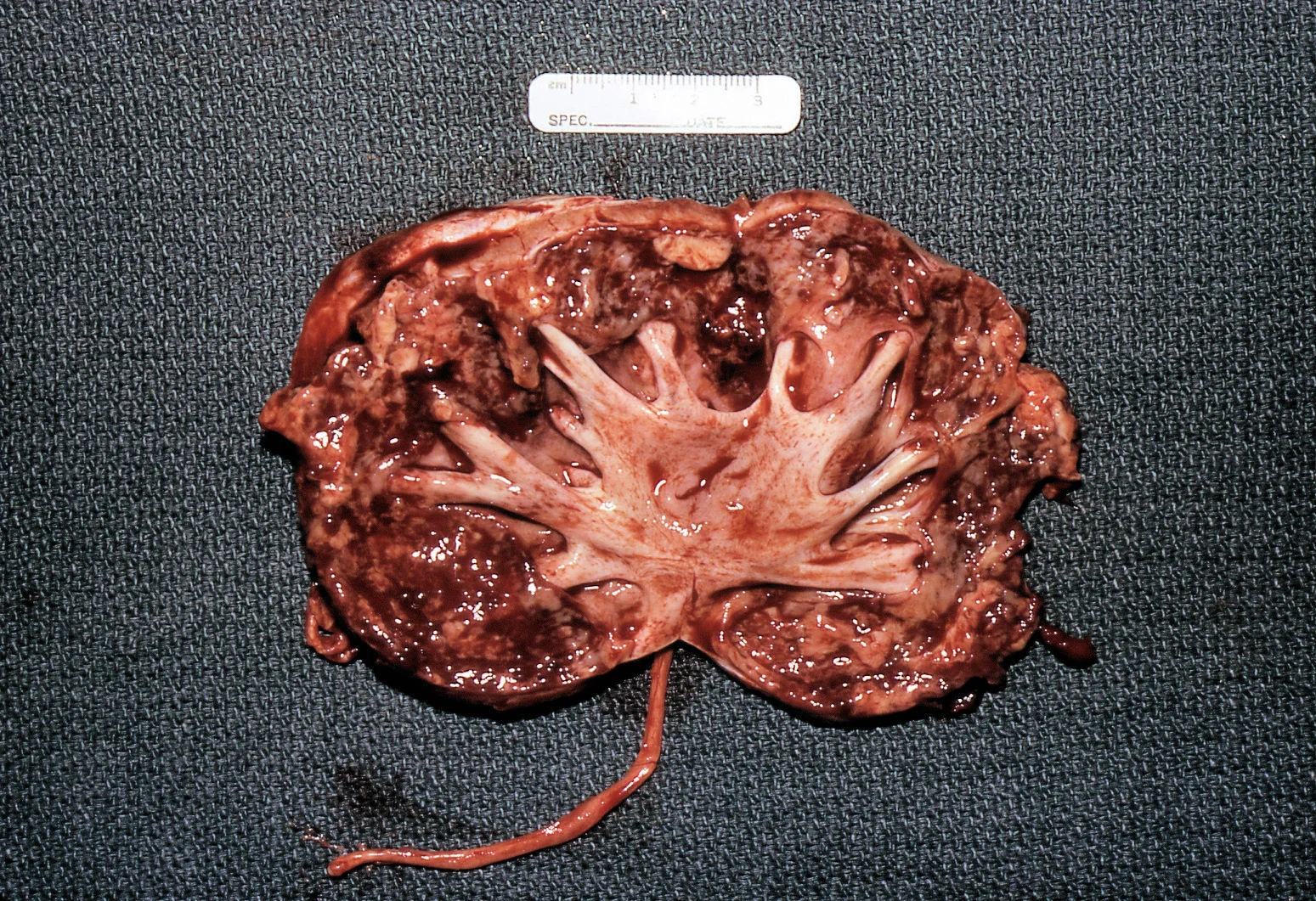
Surgical specimen from a dog showing a dilated renal pelvis and infected and necrotic parenchyma
Pyonephrosis in humans refers to infected urine in an obstructed collecting system. It can begin as pyelonephritis and then convert to pyonephrosis when outflow obstruction and hydronephrosis occurs (Figure 1). The infection can result from an ascending lower urinary tract infection or from hematogenous spread of infection from a distant primary site.
The consequences of delayed diagnosis include urosepsis, renal abscess formation, and irreversible renal damage, which might require nephrectomy. Treating pyonephrosis with antimicrobial drugs without correction of the obstruction may still be potentially devastating for the patient.
Clinical Signs
The clinical signs seen in the cases I’ve examined have varied according to the stage of severity. Early on, the patient can have bacteriuria without clinical signs. However, as the condition progresses to cause a large accumulation of pus, bacteria, and necrotic debris within the renal pelvis, the patient will eventually show signs of urosepsis consisting of fever, mental depression, decreased appetite, and eventually prostration. Variable abdominal discomfort with renomegaly is present during renal palpation.
Diagnostic Testing
Abdominal radiographs will show only an enlarged kidney or kidneys (Figure 2A), but excretory urography will show renal pelvis dilatation (Figure 2B). Ultrasonography will also show hydronephrosis, renal parenchymal disarray, and necrotic debris in the renal collecting system (Figure 3).

Abdominal radiographs of a cat with bilateral pyonephrosis; a cystotomy performed 6 months previously had caused bilateral ureteral obstruction at the urethral junction (A and B). Pyelography was done by pyelocentesis, and bilateral hydronephrosis is shown.

Abdominal radiographs of a cat with bilateral pyonephrosis; a cystotomy performed 6 months previously had caused bilateral ureteral obstruction at the urethral junction (A and B). Pyelography was done by pyelocentesis, and bilateral hydronephrosis is shown.

Abdominal ultrasonography (from patient in Figure 2) showing hydronephrosis and necrotic debris in both renal pelvises (A); close-up view of the right kidney (B).

Abdominal ultrasonography (from patient in Figure 2) showing hydronephrosis and necrotic debris in both renal pelvises (A); close-up view of the right kidney (B).
Only ultrasonography-guided fine needle pyelocentesis allows specific sampling of urine and, thus, an exact diagnosis of infection. In addition, this procedure permits microbial identification and cytologic assessment, and can also facilitate a direct injection of renal contrast medium into the renal pelvis for contrast urography.
The laboratory evaluation will often show leukocytosis, slight to moderate anemia, and azotemia if there is complete outflow obstruction. With partial obstruction, urinalysis can show overt pyuria and bacteruria; however, if the involved side is completely obstructed, the urine draining from the normal kidney may give the incorrect impression of a sterile urinary tract.
Therapy & Prognosis
Treatment entails the appropriate use of nephrostomy tube drainage or nephrectomy, along with the administration of an antimicrobial drug that is effective against the infective organism. Therefore, urine culture and sensitivity testing are essential. Potentially nephrotoxic antibiotics should not be used.
Intravenous isotonic crystalloid solutions should be given to correct dehydration and address maintenance fluid needs. Urosepsis, as defined by systemic spread of a urinary infection, should be managed under intensive care conditions. Prognosis is fair with unilateral renal involvement and more guarded with bilateral involvement.
Pyonephrosis in dogs has been described in the veterinary literature, although it is considered rare compared with pyelonephritis. My literature search identified no veterinary references to pyonephrosis in cats.