Prostate Cytology
A 7-year-old, intact male dachshund presented with a 2-month history of lethargy, decreased appetite, and tenesmus.
History. In addition to the above clinical signs, the owners had also noticed that the dog was reluctant to jump and was licking his ventrum, perineum, and hindlimbs.
Physical Examination. The dog was considerably lethargic on initial physical examination. Temperature, pulse, and respiratory rate were within normal limits. Bilateral perineal hernias were present. Pain was elicited upon palpation of the lumbosacral area. On rectal examination, the prostate was severely enlarged and adhered to the pelvic canal.
Laboratory Analysis. A hemogram did not reveal any clinically significant abnormalities. Serum biochemistry panel revealed slightly elevated ALT, but was otherwise unremarkable. Urinalysis revealed TNTC white blood cells, a moderate amount of red blood cells, and a large number of gram-negative rods. Urine culture isolated a substantial number of beta Escherichia coli.
Diagnostic Tests. Abdominal radiographs revealed a left-sided perineal hernia containing a soft tissue opacity consistent with a focal accumulation of fecal material; the urinary bladder and prostate were poorly visualized.
Abdominal ultrasonography revealed a focal area of hypoechogenicity on the cranial border of the prostate; however, due to the caudal displacement the remaining prostate could not be assessed. These changes were most consistent with prostatitis or prostate neoplasia. Three-view thoracic radiographs did not reveal any evidence of metastasis. Enlarged sternal lymph nodes indicated abdominal disease.
CT was done to further evaluate the prostate and revealed a large, nonuniformly contrast-enhancing mass causing lateral deviation of the colon. While the dog was under general anesthesia, 2 ultrasound-guided biopsy samples were taken using a 16-g, 1.7-cm throw Bard Monopty biopsy instrument needle. One of the biopsy samples was carefully rolled on a slide and submitted for cytologic examination. No hemorrhage was noted after the biopsy.

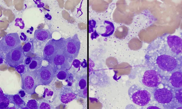
Cytologic Assessment. Cuboidal epithelial cells were seen, both in clusters and individually, with round nuclei and small amounts of pale staining cytoplasm (Figure 1). The cells had moderate anisocytosis and anisokaryosis. There were a few clusters of larger cells showing marked anisocytosis and anisokaryosis with prominent nucleoli. A moderate amount of degenerate neutrophils were present with phagocytosis of rod-shaped bacteria. The same bacteria are also seen free in the background (Figure 2).
Figure 1. Prostatitis. Note the island of cuboidal to columnar epithelial cells (prostate epithelial cells) with inflammatory cells (mainly neutrophils) in the background (arrows).
Figure 2. Note the neutrophil with intracellular rod-shaped bacteria in the upper left-hand corner (arrows ). Gram-negative bacteria, particularly E coli, are the most common isolates implicated in bacterial prostatitis.

DIAGNOSIS: BACTERIAL PROSTATITIS Prostate disease occurs most commonly in middle-aged to older, intact male dogs; however, neutered male dogs are still at risk for prostate neoplasia. Prostate diseases (benign prostatic hyperplasia [Figure 3], squamous metaplasia, bacterial prostatitis, prostatic abscess, prostatic adenocarcinoma, or transitional cell carcinoma) often present with similar clinical signs, such as hematuria, dysuria, constipation, tenesmus, and hindlimb lameness.
Figure 3. Benign prostatic hyperplasia. Note the honeycomb appearance of clusters of normal prostatic epithelial cells.
Diagnostics. When prostate disease is suspected, initial evaluation should include physical examination, rectal examination, abdominal radiography and ultrasonography, urinalysis with urine culture and susceptibility, prostate fluid analysis (culture and cytology) from prostatic wash or ejaculation, and ultimately fine-needle aspiration or biopsy for definitive diagnosis. An accurate and timely diagnosis is necessary due to the varying treatments and prognosis of different conditions.
One study found that there was strong agreement between cytologic and histopathologic diagnosis for prostate conditions.1 In this same study, cytologic assessment agreed with histopathologic diagnosis in 20 of 25 cases (80%). Normal prostate gland cytology consists of clusters of cuboidal to columnar prostatic epithelial cells that should be uniform in appearance and 10 to 15 microns in diameter. The nucleus is round to oval and located at the base of the epithelial cells. The cytoplasm should be granular and basophilic.
In cases of benign prostatic hyperplasia, cells should resemble those of a normal prostate. When inflammation is present, numerous neutrophils are usually seen in addition to macrophages, lymphocytes, and plasma cells. With bacterial prostatitis, phagocytized and free bacteria are seen and neutrophils are often vacuolated and exhibit karyolysis. Adenocarcinoma cells of the prostate are usually large, ranging in size from 20 to 80 microns in diameter. The cytoplasm is vacuolated and may contain eosinophilic material. Nuclei are large, round to oval, and multiple nucleoli are often seen. In addition, mitotic figures and multinucleate cells are often seen. Transitional cell carcinoma cells appear similar to adenocarcinoma cells.
Treatment. Chronic bacterial prostatitis requires long-term antibiotic therapy, usually for at least 4 weeks. The most common bacteria isolated are E coli and Proteus, Pseudomonas, Streptococcus, and Staphylococcus species. Antibiotic selection should be based on culture and sensitivity results and should include those agents that will reach the highest concentration in the prostate.
ASK YOURSELF ...
What methods can be used to obtain prostatic fluid for cytology and bacterial culture?
What are potential complications of fine-needle aspiration or biopsy of the prostate?
Did You Answer ...
Prostatic fluid can be obtained via ejaculation (collection of the third fraction) or by prostatic wash. In cases of acute prostatitis or prostatic abscess, prostatic wash may cause septicemia by forcing organisms into the bloodstream. Treatment with antibiotics before prostatic wash is recommended. Urine bacterial culture results often correlate with prostatic fluid culture results and may be used as an alternative if the other procedures cannot be performed.If neoplasia is suspected, fine-needle aspiration or biopsy of the prostate is recommended because neoplastic cells are rarely recovered from samples obtained by prostatic wash or ejaculation.
Hemorrhage and peritonitis can occur. Prostate biopsy is not indicated in cases of acute prostatitis unless neoplasia cannot be ruled out. Mild, transient hematuria may occur but usually does not require treatment. Ultrasound-guided biopsies are recommended over blind biopsies to reduce potential damage to surrounding structures.
PROSTATE CYTOLOGY • Brandy Dugas and Jill Brunker
Reference
1. Evaluation of the cytologic diagnosis of canine prostatic disorders.Rowe JR, Canfield PJ, Martin PA. Vet Clin Pathol 3:150-154, 2004.