Primary Glaucoma in a Siberian Husky
Shannon D. Boveland, DVM, MS, DACVO, Auburn University
A 2-year-old, 52.8-lb (24-kg), intact female Siberian husky is presented with a cloudy, painful left eye of 3 weeks’ duration. Her owner reports she has been increasingly lethargic over the past 2 weeks. Her appetite is normal, and there is no history of vomiting. Urination and defecation are also normal. The referring clinician previously prescribed bacitracin/neomycin/polymyxin B/dexamethasone (BNP/DEX) ointment every 8 hours for 1 week for her left eye and cephalexin 20 mg/kg PO every 12 hours for an ear infection.
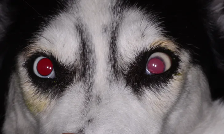
Ophthalmic examination of the left eye reveals severe blepharospasm and episcleral injection with mild epiphora. Negative direct and indirect pupillary light reflexes as well as negative dazzle and menace reflexes are observed. Schirmer tear test is 13 mm/minute (normal, 15-25 mm/minute), and intraocular pressure (IOP) is 43 mm Hg (normal, 15-25 mm Hg). Mild focal corneal edema is noted with 360-degree neovascularization. The pupil is mydriatic and unresponsive with mild color change in the iris indicative of inflammation (Figure 1). Fundic examination reveals cupping of the optic nerve head (Figure 2).
Findings in the right eye are within normal limits; Shirmer tear test is 24 mm/minute, and IOP is 15 mm Hg. Gonioscopy reveals an abnormal (narrow-closed) iridocorneal angle. Menace, dazzle, and direct pupillary light reflexes are positive, and indirect pupillary light reflex is negative.
Based on ophthalmic findings, primary glaucoma with secondary uveitis is diagnosed. Remainder of the physical examination is unremarkable.

Mild focal corneal edema, 360-degree limbal neovascularization, mydriatic pupil, mild color change in the iris (due to secondary uveitis), conjunctival hyperemia, and a slightly elevated third eyelid can be seen in the patient’s left eye. Image courtesy of Auburn University

Fundic image of the patient’s left eye. Cupping of the optic nerve head, moderate vascular attenuation of retinal vessels, and depigmentation of the nontapetum can be seen. Image courtesy of Auburn University
Take Our Quiz
Conclusion
A single glaucoma medication does not typically lower or maintain IOP in the normal range. For acute primary glaucoma, emergency medical treatment should consist of a combination of drugs that decrease production and increase outflow of aqueous humor as well as dehydrate the vitreous and anterior segment spaces.
CAI = carbonic anhydrase inhibitor, IOP = intraocular pressure, POAG = primary open-angle glaucoma