Practical Guide to Tick-Borne Disease
Profile
Definition
Tick-borne diseases are caused by etiologic agents that are transmitted from ticks to dogs; such agents include Ehrlichia, Anaplasma, Babesia, Rickettsia, hemotropic Mycoplasma, Francisella, Hepatozoon americanum, and Borrelia species.
Systems
Ticks cause multisystemic disease in dogs.
Hematologic changes are common and sometimes typical (e.g., thrombocytopenia)
Multiorgan dysfunction with associated clinical signs and clinicopathologic changes are common (Ehrlichia, Anaplasma, Rickettsia, H. americanum).
Single organ dysfunction can occur (arthropathy in borreliosis).
Incidence/Prevalence/Geographic Distribution
Incidence and geographic distribution are dependent on distribution of the tick vector throughout the United States (Table 1).
Prevalence within a geographic region is usually dependent on season and ecology of the tick vector (Table 1).
Breed Predilection
All dogs are susceptible to tick-borne disease.
Babesia gibsoni has shown an increased incidence of infection in American Staffordshire and American pit bull terriers; disease is believed to be acquired during fighting.
German shepherds and Doberman pinschers develop more chronic and severe forms of rickettsial diseases.
Risk Factors
Major factor is exposure to ticks (young hunting dogs with extensive outdoor activity).
Exposure depends on ecology of the tick, including the season during which it is most prevalent, infection rate of the disease in the tick species, attachment requirements of tick to transmit disease-American dog tick requires attachment > 5 hours to transmit R. rickettsii; ticks of the Ixodes species require 24 hours of attachment to transmit Borrelia burgdorferi; Amblyomma maculatum requires ingestion to transmit H. americanum.
Blood transfusion-receiving blood from unscreened donors from enzootic regions is a high risk factor.
Splenectomy predisposes dogs to clinical disease with M. hemocanis; a milder form of E. canis infection also occurs in splenectomized dogs.
Concomitant infection with tick-borne or other diseases or a weakened immune system (from age, immunosuppressive drug therapy, or congenital defects) predisposes to more severe disease.
Pathophysiology
Depends on the disease.
Rickettsials-Vasculitis and immune mechanisms lead to endothelial damage in multiple organs, thrombocytopenia, hyperglobulinemia, and eventual bone marrow depletion with pancytopenia. Different organisms manifest with different organ involvement-for example, E. ewingii mainly centers on joints and the central nervous system, A. platys mainly affects platelets, R. rickettsii mainly affects endothelial cells of small arteries and venules. Incubation period is 8 to 20 days.
Babesia-Intravascular and extravascular hemolysis with anemia. Transplacental transmission occurs.
Mycoplasma-Splenectomy, immunosuppression from concomitant infectious diseases, or drug therapy can precipitate anemia in which immune mechanisms are also involved. Transplacental transmission does not occur.
Francisella-Macrophages become infected and spread the organism to lungs, spleen, liver, lymph nodes, and skin where microabscess formation occurs.
Borrelia-Few infected dogs develop clinical signs; immune mechanisms against borrelial proteins and cytokine responses are implicated in pathologic conditions of the joint. Transplacental transmission rare but can occur.
Hepatozoon-Infected ticks are ingested; sporozoites enter blood and lymphatics via stomach mucosa then target lymphatic tissue, bone marrow, or other organs (liver, lungs, kidneys), causing hepatitis, pneumonitis, and glomerulonephritis. Transplacental transmission occurs.
Signs
There is no one clinical sign typical of tick-borne diseases (Table 2).
Travel history should be carefully considered (Table 1).
Many infections, such as those caused by Borrelia, Babesia, Rickettsia, Anaplasma, and Mycoplasma, are asymptomatic, but may manifest with clinical signs if they occur concomitantly with other infections or immunosuppression.
Clinical signs usually indicate multisystemic involvement, which should always prompt the clinician to place tick-borne diseases high on the differential diagnosis list.
Some tick-borne diseases do show more specific signs, such as severe debilitation, intermittent antibiotic-nonresponsive fever, weight loss, muscle atrophy, and hyperesthesia; mucopurulent ocular discharge occurs in most dogs infected with H. americanum.
Diagnosis
Identification of a tick removed from a patient with specific clinical signs can provide direction in diagnosing the specific pathogen (Table 1, see below for F**igures**).
Clinical signs should be correlated within the geographic area in which the patient resides or has traveled. For example, a dog from New Jersey that presents with joint pain (polyarthritis) is likely to have borreliosis; a dog from Oklahoma with joint pain probably has E. ewingii infection.
Important and common clinical signs often associated with tick-borne diseases include fever, chronic weight loss, petechiae/ecchymoses, and lymphadenopathy.
Clinicopathologic findings (Table 2) in conjunction with clinical signs should prompt a more specific search (serologic evaluation) for certain pathogens.
Important and common clinicopathologic findings that should trigger the clinician to think of tick-borne diseases include thrombocytopenia (rickettsial diseases, Babesia, Francisella), regenerative anemia (Babesia, Mycoplasma), hyperglobulinemia (Ehrlichia, H. americanum), and profound leukocytosis (H. americanum).
Many tick-borne diseases cause visible intracellular inclusions that, although rare for some agents, are definitively diagnostic if found:
Ehrlichia—Morulae within leukocytes (E. canis, E. chaffeensis, E. ewingii, A. phagocytophilum) or platelets (A. platys).
Babesia—Large, often in doubles, piriform organisms (B. canis) or smaller, single organisms (B. gibsoni) within red blood cells.
Mycoplasma—Chains of organisms on red blood cell surfaces.
H. americanum—Gamonts within neutrophils or monocytes.
Tick-borne diseases can occur concomitantly, so use serologic testing and other pathogen-specific assays to search widely; the finding of one disease should prompt a search for others transmitted by the same tick species or others that are enzootic to a specific geographic area.
Results of serologic tests need to be assessed in the context of prevalence, which affects the predictive value of a test.
Cross-reaction with other pathogens is often common, especially within the rickettsial group (Table 3).

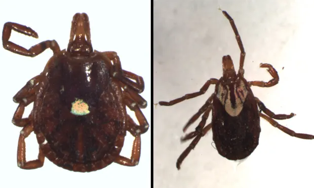
Figure 1.
Unengorged female of Amblyomma americanum (bar = 1 mm)
Treatment
Most dogs can be treated as outpatients.
The sooner treatment is initiated, the more favorable will be the prognosis.
Dogs with chronic tick-borne diseases are rarely curable.
Supportive therapy with fluids for dehydration (rickettsial diseases, Francisella), and blood transfusion (Babesia, Mycoplasma) may be indicated but will not appreciably increase platelet or leukocyte counts.
Medications
Although there is a range of drugs for rickettsial agents, doxycycline is the most commonly used agent (Tx at a Glance).
Imidocarb may be a first choice for rickettsial diseases as well at Babesia canis if a definitive diagnosis has not yet been made.
Whether dogs that are serologically positive for Borrelia but clinically normal should be treated with doxycycline or other antibiotics is controversial. Because fewer than 5% of Borrelia-positive dogs ever develop clinical signs and treatment does not guarantee removal of the organism, there seems to be little rationale for antibiotic treatment in these dogs at this time.
Early use of glucocorticoids in diseases with an immune-mediated component (rickettsial diseases, Babesia, Mycoplasma) may improve the outcome as long as they are used in conjunction with appropriate antibiotic therapy.
Glucocorticoids used for immunosuppression in suspected cases of immune-mediated thrombocytopenia will not negatively affect concurrent treatment of rickettsial agents.
Follow-up
Response to therapy during acute stages of disease caused by rickettsial agents, Babesia, Mycoplasma, and Borrelia should occur within 24 to 48 hours.
Most follow-up should consist of physical examination, CBC, biochemistry panel (globulins, liver enzymes, renal function), and urinalysis rather than serologic testing.
Platelet counts should return to normal within 5 to 8 days. If not, seek another cause.
Repeat platelet counts at the end of therapy and again in 6 months.
Serologic testing and blood polymerase chain reaction are not effective methods of assessing the efficacy of therapy with ehrlichial diseases; reinfection is possible.
Long-term immunity does develop after successful treatment of R. rickettsii.
Therapy is seldom effective in removing the organism in most cases of tick-borne disease, so lifelong monitoring for return of clinical signs is essential. The exception is that a single dose of imidocarb is believed to eliminate the carrier state of B. canis.
Prevention
Vaccines are only available for Borrelia and should only be used in enzootic areas.
Whole-cell vaccines and recombinant OspA vaccines induce antiOspA antibodies to develop in the host; these antibodies are passed to the tick while feeding and incapacitate the Borrelia organisms within the tick, thereby preventing transmission.
Vaccination of an already infected dog is not indicated.
Tick control is essential.
Effective products include fipronil (Frontline-Merial), imidocloprid/ permethrin combination (K9 Advantix-Bayer), and amitraz (Preventic-Virbac). Use of spot-on or oral flea control products combined with pyrethrum-based collars is also effective.
Daily body checks for attached ticks is likely to be effective in preventing infection with tick-borne diseases, although it requires considerable effort for the owner, who must wear gloves and use tweezers to remove the ticks. Ticks need to be disposed of carefully (incinerated) to prevent dogs from eating them.
Some have advocated the use of tetracycline (6.6 mg/kg, PO Q 24 H) as chemoprophylaxis against the rickettsial agents, Borrelia, and Mycoplasma, for dogs entering enzootic areas, although this approach may not be practical.
Screening of blood donors or blood used for transfusion and limiting fighting behavior in pit bull terriers are important.
Prognosis/Future Considerations
It is often difficult to separate recrudescence from reinfection in most tick-borne diseases.
Response to therapy for cases of definitively diagnosed rickettsial agents can be expected to be approximately 70%.
All Borrelia cases showing clinical signs of polyarthropathy should improve dramatically after 3 days of antibiotics-failure to do so should prompt further investigation of the joints for other causes, especially immune-mediated.
Dogs treated for babesiosis clinically improve within 24 hours but few drugs eliminate the parasites. However, because of the balance between low parasite numbers and host immune response, hemolytic crises during recurring infection are rare.
The long-term prognosis for dogs infected with H. americanum is good (Tx at a Glance). Relapses do occur, but a 2-year survival rate of 84% can be expected. Some dogs live for more than 5 years after diagnosis. Death usually results from glomerulonephritis and amyloidosis.
Few reports of diagnosed Francisella or survival of Francisella have been documented in dogs.
Tx at a Glance
TMS = trimethoprim-sulfadiazine