Polyuria, Polydipsia, & Hypercalcemia
A 10-year-old spayed female mixed-breed dog was evaluated for polyuria/polydipsia. A palpable mass was found in the thyroid region. The intact parathyroid hormone was 128.5 (reference range, 27-155), ionized calcium 1.63 (reference range, 1.24-1.47), and total calcium 13.2 (reference range, 8.9-11.4). Ultrasonography of the thyroid region found 3 circular hypoechoic structures approximately 4 mm in diameter. An abdominal ultrasound was performed to rule out other causes of hypercalcemia, such as lymphoma, and, surprisingly, both adrenal glands were enlarged. It is believed that the clinical signs and hypercalcemia were a result of primary hyperparathyroidism.
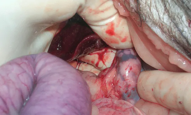
Three masses were removed from the left thyroid region during 2 separate surgeries. The right cranial parathyroid was also enlarged but not removed. All 3 masses were parathyroid adenomas; at necropsy 11 months later an adenoma was found in the right cranial parathyroid. Both enlarged adrenals had adenocarcinomas, with the right side partially seeded in the vena cava. Figure 1 shows the excised left adrenal gland (7 cm long); Figure 2 shows the right adrenal gland seen inside the caudal vena cava. The dog was euthanized due to excessive bleeding from an oral malignant melanoma.