Polyarthritis in a Dog
John W. Harvey, DVM, PhD, DACVP (Clinical Pathology), University of Florida

A 3-year-old, male Labrador retriever was presented with reluctance to move.
History
The dog began having difficulty getting in and out of the car 5 days previously. He also became lethargic and febrile and had a decreased appetite. Aside from thrombocytopenia, results from the CBC and clinical chemistry panel were within normal limits. The dog was treated with a glucocorticoid and intravenous lactated Ringer's solution containing cefazolin by the referring veterinarian. When clinical signs did not improve, the dog was referred to the University of Florida Veterinary Medical Teaching Hospital for evaluation.
Physical Examination
Findings included depression, weight loss, stiffness in the hind limbs, swollen stifle and hock joints, enlarged prescapular and popliteal lymph nodes, hair loss preceding the current clinical presentation, and tapeworms.
Table: Laboratory Findings*
* Routine culture of joint fluid was negative; serologic tests were negative for Rickettsia rickettsii, Borrelia burgdorferi, and antinuclear antibody. Other findings on the CBC, serum chemistry, and urinalysis were unremarkable.

FIGURE 1
Ask Yourself ...
Was this dog infected with Ehrlichia canis?
Could it have been infected with more than one rickettsial organism?
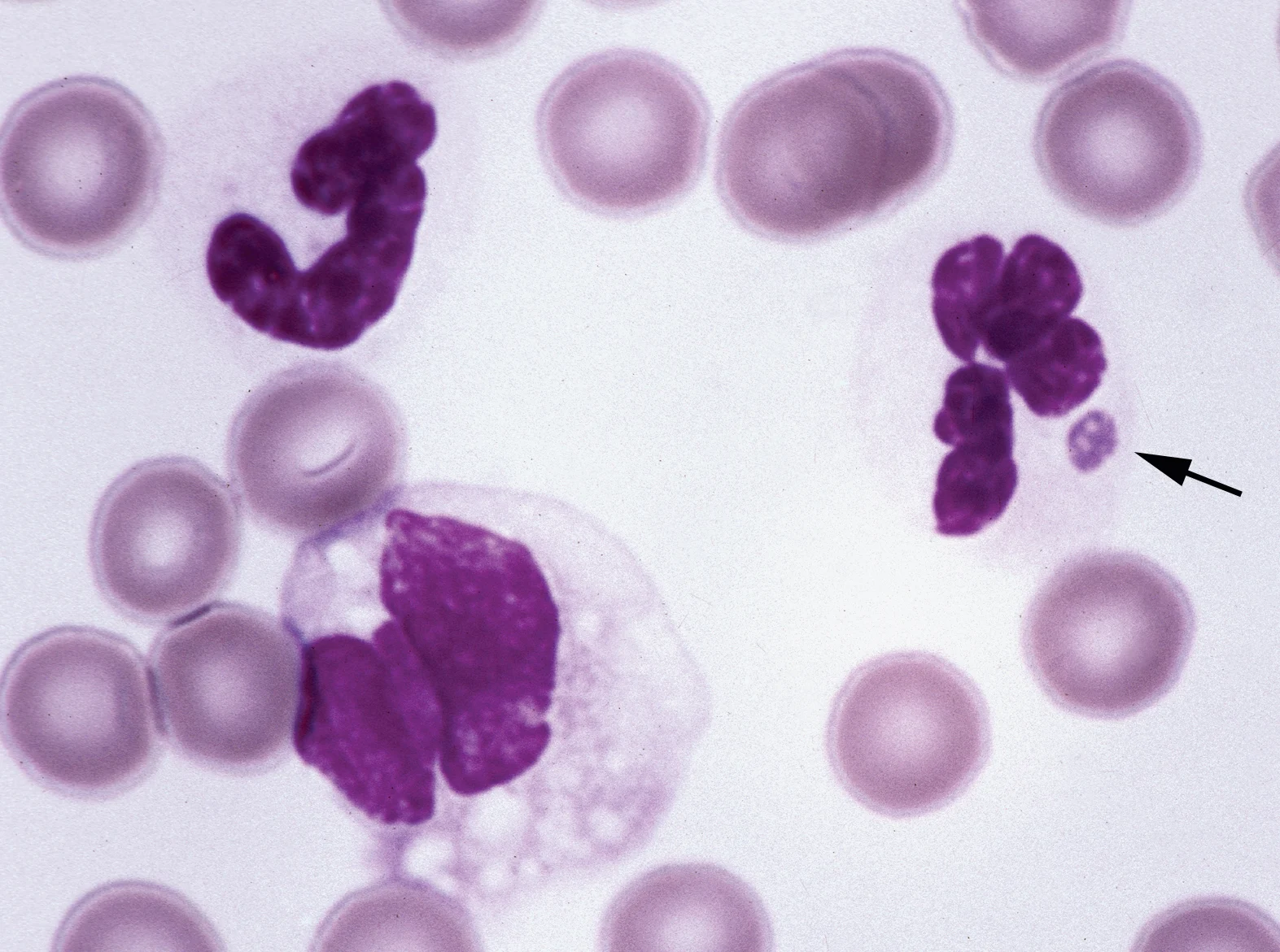
What is the most likely organism reported to infect dogs that would account for the polyarthritis and neutrophilic inclusions?
What additional diagnostic tests might definitively identify the causative agents in this case?
Serum and blood samples were submitted to the Centers for Disease Control and Prevention for further evaluation. Repeated testing by CDC revealed high serum IFA titers for both E. canis (1:4096 and 1:8192) and E. chaffeensis (1:8192 and 16,384). E. chaffeensis was initially recognized to cause disease in humans, but it can also infect dogs. Attempts were made to amplify Ehrlichia DNA for the 16S rRNA gene from blood using a nested PCR with a specific panel of primers. Neither E. canis nor E. chaffeensis could be identified, but a primer specific for E. ewingii generated a 300 base pair segment that when sequenced was identical to that published for E. ewingii.
Diagnosis: Ehrlichia ewingii infection

FIGURE 1
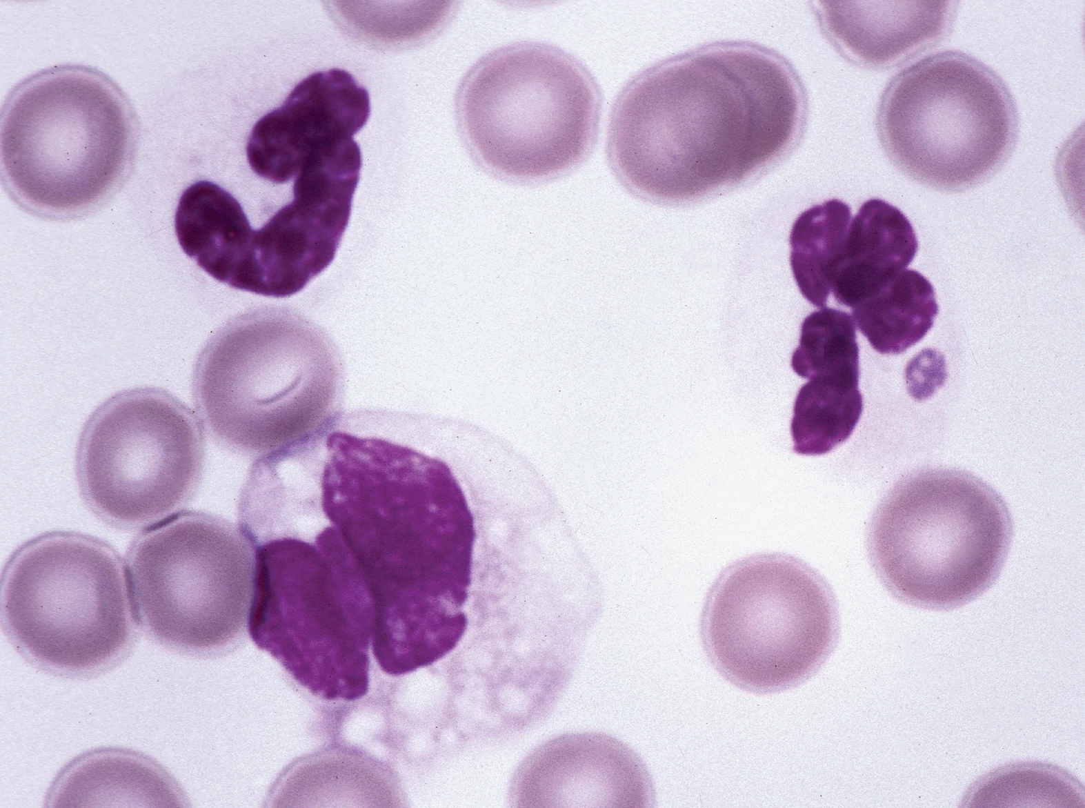
Blood film containing two neutrophils and a monocyte. A cytoplasmic inclusion is present in one neutrophil (arrow) with the structure of a morula typically seen in rickettsial infections. (Wright-Giemsa stain)
Did You Answer ...
Possibly, but not necessarily. Serologic cross-reactivity occurs between genetically related rickettsial organisms. Ehrlichia canis and E. chaffeensis are especially closely related organisms that cannot be differentiated using conventional IFA tests. Although the highest titers were for these organisms, it is difficult to attribute the presentation in this case solely to E. canis or E. chaffeensis infections, because both agents infect mononuclear phagocytes, and morulae were seen in neutrophils.
Probably. Dogs and tick vectors that spread rickettsial disease may be infected with several rickettsial species (as well as other infectious organisms) simultaneously. Although E. canis and E. chaffeensis were not identified by PCR, the IFA titers determined by the Centers for Disease Control are higher than one might expect for a cross-reaction with E. ewingii. Consequently, this dog could also be chronically infected with E. canis or E. chaffeensis but not have sufficient numbers of organisms in blood to be identified by PCR.
Although arthritis has been reported in dogs believed to have been infected with E. canis, this diagnosis was based on serum IFA tests and not on genetic identification of the organism involved. Infections with other rickettsial agents, including E. ewingii, might also result in a positive E. canis titer, or the dogs might have had concurrent infections with more than one organism. Morulae have been identified in neutrophils of dogs infected with two rickettsial organisms, A. phagocytophilum (formerly E. equi [horses], E. phagocytophila [ruminants], and human granulocytic ehrlichiosis agent) and E. ewingii. Polyarthritis has been associated with E. ewingii infections, but not with A. phagocytophilum infections. Based on these considerations, an E. ewingii infection was suspected, but a specific serologic assay for E. ewingii was not available.
PCR-based assays that can identify specific infectious organisms based on their genetic composition. The 16S rRNA gene is currently the most widely studied gene for bacterial identification. In this case, E. ewingii infection was proven by sequencing a portion of the 16S rRNA gene amplified by PCR.