Phalangeal Fillet Technique for Digital Pad Transfer
Nicole Ehrhart, VMD, MS, Colorado State University
Footpads, which are highly specialized epithelial and stromal tissue structures, consist of a shock-absorbing, fatty, elastic, collagenous cushion covered by a tough, antiskid, epithelial surface. When injured, footpads heal slowly because they are under shear and tension stress during ambulation.
Footpad Loss
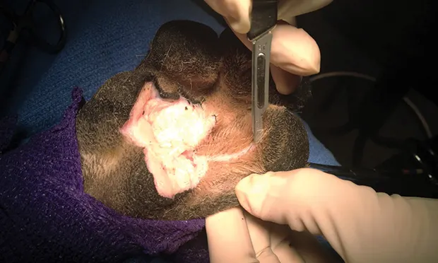
Loss of the major weight-bearing footpads—metatarsal (MT) and metacarpal (MC)—may result in permanent lameness, especially in large dogs. Partial or complete loss can result from extensive traumatic tissue damage or tumor resection (Figure 1). No other cutaneous tissue can completely serve as a substitute for this highly specialized, weight-bearing surface.

Large metacarpal pad melanoma in which extensive resection would be necessary to achieve adequate tumor control. The phalangeal fillet technique could be used to reconstruct the metacarpal pad in this case.
With extensive MC or MT footpad loss, healthy digital pads, with a surface identical to the original pad tissue, can be transferred for reconstruction. The phalangeal fillet technique involves transferring one or two digital pads on a vascularized cutaneous pedicle to the area of the missing MC or MT footpad.1
The outer digits (phalange [P]2 and/or P5) are most commonly used, but any of the 4 can be used (Figure 2). If a second digital pad is needed for additional weight-bearing surface, the procedure can be repeated on an additional digit. The opposing edges of adjacent pads should be trimmed to provide fresh edges for appositional healing.

Before digital pad transfer in a cadaver limb from a 70-kg mastiff with partial metacarpal pad resection.

After digital pad transfer in a cadaver limb from a 70-kg mastiff with partial metacarpal pad resection.
Postprocedural Healing
After the procedure, a heavily padded bandage and external support (eg, Mason Meta Splint, jorvet.com) must be applied to the limb. The bandage should be changed q3–4d for 3 weeks until healing is complete. Exercise should be restricted for 4 weeks for optimal healing.
Some rigid support to the bandage is essential to transmit the weight-bearing forces through the splint and not onto the digital pads. The rigid support should be on the palmar or plantar surface and extend just beyond the digits so the splint (ie, not the digits) hits the ground during ambulation. If the paw contacts the ground, then the forces transmitted during weight bearing can inhibit healing or lead to flap failure.
Complications & Outcome
Partial dehiscence of the digital pad is not uncommon, especially if patient activity is unrestricted or the owners are noncompliant with bandage changes. If partial dehiscence occurs, sutures may be replaced after gentle cleansing and debridement. Exercise restriction should be maintained and splinted bandages worn for longer than 4 weeks, if needed. With patience, the paw pads usually heal following this procedure, and patient function is typically good.
Related Article: Pressure-Related Wounds: Prevention & Treatment
Related Article: Problem Wound
Step-by-Step: Phalangeal Fillet Technique for Digital Pad Transfer
What You Will Need
Standard surgical instrument pack
Esmarch bandage (tourniquet)
#15 scalpel blade
Sterile self-adhering elastic support wrap (eg, Vetrap or Coban, 3m.com)
Bandage material
Splint material

STEP 1A
Clip the fur from the distal limb and prepare the affected foot for surgery. To diminish the bacterial load on the skin, soak the paw for 5 minutes in an examination glove filled with 0.12% chlorhexidine diacetate surgical preparation solution diluted with tap water before standard surgical skin preparation.