Peripheral Nerve Block Techniques: Dental Blocks
Janet A. Welch, DVM, Diplomate ACVS, Auburn University
Overview
Peripheral nerve blocks are performed by injecting local anesthetic in the immediate vicinity of a peripheral nerve or nerve plexus. Common peripheral nerve blocks in small animal patients include epidural, brachial plexus, forefoot, intraarticular stifle, and dental blocks. All but dental peripheral nerve blocks were covered in the March 2004 issue, pages 47-50.
Local anesthetic blocks can be given to heavily sedated or anesthetized animals. Greatest efficacy is obtained when they are given before surgery is begun. Effective peripheral nerve blocks provide excellent anesthesia and analgesia and can reduce the amount of inhalation anesthetic needed to perform a procedure as well as mitigate the "wind-up" response of the CNS to noxious stimuli.
The duration of effect varies with the type of local anesthetic. (See March, page 47, for further information about specific local anesthetic agents and tips about their use.) Shorter-acting agents provide 1 hour of anesthesia, and longer-acting agents provide up to 6 hours. The duration of analgesia, or reduced sensation of pain, may be longer.
Opioid analgesics, such as morphine, can be administered locally along with lidocaine or bupivacaine to augment the duration of analgesia. In addition to the CNS, opioid receptors are present in the periphery if tissues are inflamed.
Applications & Necessities
Sensory nerve fibers that innervate the bone, teeth, and soft tissues of the oral cavity emanate from the maxillary or mandibular branches of the trigeminal nerve. Four regional nerve blocks can be readily performed to provide analgesia for dental and oral surgical procedures.
Related Article: To Extract or Not to Extract
Related Article: Surgical Extraction: Maxillary 4th Premolar Tooth in a Dog
Materials
Tuberculin syringe
25- to 27-gauge, 3/4- to 1-inch needle
Drugs and Dosages
Bupivacaine 0.5%: 0.1 to 0.5 ml per site for dogs and 0.1 to 0.3 ml for cats. An additional 50% of the agent can be administered for an infraorbital nerve block. The peak effect occurs within 30 minutes and the duration is 4 to 6 hours.
Do not exceed a maximum cumulative dose of 2.0 mg/kg.
Step-by-Step: Mental Nerve Block
The mental nerve block anesthetizes all oral tissues rostral to the second premolar on the infiltrated side. The middle mental foramen is the largest of the three and is used for this block.
Palpate the mental foramen just ventral to the rostral (mesial) root of the second premolar.
Advance the needle into the opening of the foramen in a rostral-to-caudal direction and slowly inject.
Hold digital pressure over the injection site for 30 to 60 seconds to ensure maximal caudal diffusion into the mandibular canal.
In cats, the foramen usually cannot be palpated. Place the needle into the submucosa at the rostral border of the labial frenulum halfway between the dorsal and ventral borders of the mandible and inject.


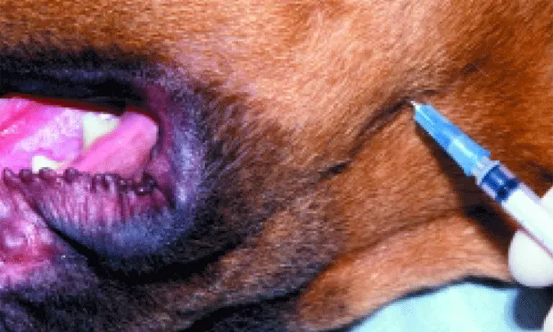
Step-by-Step: Inferior Alveolar (Mandibular) Nerve Block
The mandibular nerve block affects the bone, teeth, soft tissue, and tongue on the infiltrated side.
In dogs, there is a palpable notch on the caudal ventral mandible just cranial to the angular process. If the notch cannot be palpated, select the point on the ventral mandible that is located on a vertical plane with the lateral canthus.
Using this landmark, insert the needle at the lingual aspect of the ventral mandible.
Advance the needle to the midpoint between the ventral and dorsal borders of the mandible.
Aspirate and slowly inject.


Step-by-Step: Infraorbital Nerve Block
The infraorbital nerve block reliably anesthetizes the bone, soft tissue, and dentition rostral to, but not including, the upper fourth premolar. An additional 50% of the anesthetic agent is used for this block.
Palpate the infraorbital foramen in the maxilla dorsal to the caudal (distal) root of the upper third premolar. In some cases, this is easier to palpate extraorally.
Insert the needle through the submucosa in a caudal direction.
Advance the needle to the entrance of the foramen, aspirate, and inject while holding digital pressure over the foramen.
Withdraw the needle and hold digital pressure over the canal for 30 to 60 seconds.


Step-by-Step: Maxillary Nerve Block
The maxillary nerve block affects the bone, teeth, soft tissue, and palatal tissue on the infiltrated side.
With the patient's mouth open, palpate the notch where the zygomatic arch meets the bone surrounding the last maxillary molar.
Insert the needle directly adjacent to the bone at this level.
Hold the needle perpendicular to the hard palate.
Advance the needle dorsally to a level just beyond the extent of the root tips of the last molar.
Aspirate and slowly inject.

