Perioperative Management of Brachycephalic Dogs
Jonathan Miller, DVM, MS, DACVS (Small Animal), Eclipse Specialty & Emergency Pet Care, Whippany, New Jersey
Kristi Gannon, DVM, DACVECC, Oradell Animal Hospital
What should I consider before anesthesia and surgical correction of canine patients with brachycephalic airway syndrome?
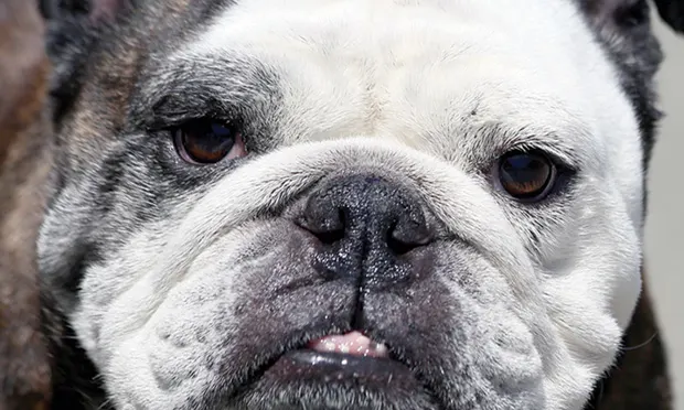
Brachycephalic dog breeds, particularly English bulldogs and pugs,1 are at increased risk for airway-related complications in the perioperative period, with death attributable to respiratory disease being the number one killer of bulldogs.2 Airflow obstruction in brachycephalic dogs can result from an elongated soft palate, stenotic nares, edematous tonsils, laryngeal edema, everted laryngeal saccules, laryngeal collapse, nasal turbinates protruding into the pharynx, edema of the epiglottis, and hypoplastic trachea.3-5 Knowledge of these airway disease components allows for careful perioperative upper airway management and greatly reduces morbidity and mortality.
Identification of High-Risk Patients
An in-depth patient history, including previous anesthesia complications and information about exercise and heat tolerance, is required to determine the relative risk for individual patients. Excitement often intensifies inspiratory difficulty and noise associated with breathing. Snoring is likely to be present in most brachycephalic breeds; however, a history of coughing, sleep apnea, or cyanosis may be predictive of perioperative risk. An obstructive breathing pattern can be recognized by slow inspiratory and abrupt expiratory phases with abducted forelimbs.
In addition, recent-onset dysphagia, vomiting, or regurgitation can suggest a loss of normal physiologic separation of swallowing and breathing mechanisms and identify patients at risk for aspiration pneumonia. The prevalence of GI disease (erosive esophageal lesions, gastritis, duodenitis) in dogs with brachycephalic airway syndrome has recently been reported as 98% with a clear correlation between severe respiratory signs and severe GI signs.6 Dogs with a history of regurgitation or vomiting should be auscultated carefully with thoracic radiographs obtained to assess for existing aspiration pneumonia, bronchial disease, hiatal hernia, or neoplasia.
Obesity plays a large role in breathing obstruction in humans.7,8 Although rarely reported in dogs with brachycephalic airway syndrome, further investigation into the role of obesity in dogs with airway obstruction is needed.
Age may also play a role in risk for postoperative complications. One study reported that dogs older than 1 year of age had a less desirable owner-assessed outcome than younger dogs.9 Age has not been investigated thoroughly as a risk factor, but suspicion should be high for perioperative breathing complications in a geriatric dog presenting with an acute exacerbation of respiratory difficulty.
The mortality rate following surgical correction of brachycephalic airway syndrome is relatively low (0%–5%), suggesting that the large majority of these dogs recover from surgery successfully.9-12 Documented mortality was often related to aspiration pneumonia or upper airway swelling. Severe laryngeal/pharyngeal edema can lead to complete airway obstruction, necessitating tracheostomy to bypass the affected portion of the airway. Identifying those dogs with preoperative exercise intolerance, cyanosis, syncope, or other recent evidence of respiratory decline may help determine which patients are at high risk for requiring a tracheostomy.
Other comorbidities should be ruled out by a CBC, serum biochemistry profile, urinalysis, and thoracic and abdominal imaging; arterial blood gas analysis or pulse oximetry can be used to identify patients with impaired respiratory function.
Preoperative Management
Patient Assessment
Following a thorough physical examination, preoperative thoracic radiographs can be pursued. Positioning for radiographs can cause a decline in respiratory function, so careful monitoring and supplemental oxygen may be necessary. Dorsoventral positioning is recommended to avoid airway collapse and patient struggling in a ventrodorsal position. The lateral radiograph should be obtained quickly with the dog immediately returned to sternal recumbency.

Radiograph demonstrating hypoplastic trachea in a bulldog puppy. The thoracic inlet (red line) and tracheal diameter (blue line) are shown.
Radiographs should be evaluated for the presence of hypoplastic trachea (defined as a ratio of tracheal diameter to thoracic inlet height of less than 0.13 for bulldogs and 0.16 for other brachycephalic breeds; Figure 1),13 bronchial collapse, bronchiectasis, pneumonia, pulmonary edema, cardiac chamber dilation, and hiatal hernia (rare). Lung assessment can be challenging because of the dorsoventrally compressed anatomy and difficulty in obtaining a true inspiratory film.
Dogs presenting in respiratory distress should be supplemented with oxygen immediately via face mask or oxygen cage. Limited handling in a quiet environment is advised. Sedation with acepromazine (0.02–0.05 mg/kg IV or IM), butorphanol (0.1–0.3 mg/kg IV or IM), or dexmedetomidine (2–5 µg/kg IV or IM) will alleviate anxiety and reduce respiratory effort, which in turn will decrease airway swelling and collapse. Monitoring body temperature will dictate the need for cooling efforts. Thoracic radiographs and blood samples (for anesthesia assessment and blood gas analysis) can be obtained after patient stabilization.
Assessing a patient’s need for oxygen, response to oxygen therapy, and need for intervention with positive-pressure ventilation and/or temporary tracheostomy is accomplished by serial patient evaluations, pulse oximetry, and arterial blood gas analysis. Oxygen supplementation should be administered when there is clinical evidence of respiratory distress.14 If signs worsen or do not improve within a few minutes of sedation and oxygen supplementation, induction and intubation to bypass the upper airway obstruction should be performed to reduce the risk for cardiopulmonary arrest.
Premedication & Anesthesia Induction
Corticosteroids are used routinely in the preoperative treatment of brachycephalic dogs (eg, dexamethasone 0.05–0.2 mg/kg IV or IM, methylprednisolone sodium succinate 1 mg/kg IV or IM), although evidence of efficacy is lacking. In theory, glucocorticoid therapy should reduce iatrogenic tissue swelling and postoperative inflammation. In one study, dexamethasone was shown to reduce post-intubation stridor following endotracheal intubation in humans, but it had no effect on re-intubation rate.15 However, another study in humans showed that methylprednisolone did reduce laryngeal edema and lower the reintubation rate.16 Much could be gained from prospectively documenting the effects of perioperative corticosteroids in dogs.
The use of anticholinergics (eg, atropine, glycopyrrolate) must be considered carefully. The benefits of increased heart rate, decreased airway secretions, bronchodilation, and reduced saliva production can be outweighed by such negative side effects as tachyarrhythmias, decreased myocardial perfusion, reduced GI motility, and decreased esophageal sphincter pressure allowing for potential reflux and esophagitis.17
Dysphagia, hypersalivation, esophageal abnormalities, and vomiting can all predispose to aspiration pneumonia. Two recent studies highlight the results of GI endoscopic examination in brachycephalic dogs.6,11 Among 73 brachycephalic dogs referred for upper respiratory signs, 98% had GI abnormalities (upper GI inflammation in 89%, gastroesophageal reflux in 32%).6 An endoscopic examination helped to dictate treatment of occult GI disease with antacids and prokinetics.11 The authors found that 80% of the GI signs resolved following surgical airway and medical treatment.11 Consideration can be given to preoperative use of prokinetics, such as metoclopramide and cisapride, as well as antiemetics.
Avoiding the use of nausea-inducing sedatives in the mu-opioid family (morphine, hydromorphone, oxymorphone) may be reasonable. Buprenorphine (0.01–0.02 mg/kg IV or IM), butorphanol (0.1–0.3 mg/kg IV or IM), or methadone (0.1–0.2 mg/kg IV or IM) may be safer choices for premedication. Acepromazine, dexmedetomidine, diazepam, or midazolam can be useful for further sedation prior to anesthesia. Preoxygenation for at least 3 minutes is recommended before induction to reduce hypoxemia during laryngeal examination and intubation. Careful monitoring for oversedation and respiratory depression during this period is important to allow for rapid intubation if necessary. IV induction with propofol (6 mg/kg) or ketamine (2–5 mg/kg IV), along with diazepam or midazolam, allows for a thorough upper airway examination and intubation for anesthesia.
Airway Evaluation


A. The typical appearance of a bulldog larynx. Note the relatively small larynx located deep within the oral cavity. B. Line drawing illustrating important laryngeal structures.
Visual examination of the upper airway should begin with evaluation of the nares for evidence of stenosis. Thorough evaluation of the pharyngeal airway structures requires sedation. Use of a handheld laryngoscope and tongue depressors for additional retraction of tissues will usually allow for adequate evaluation of the airway (Figure 2). The soft palate should not extend beyond the tip of the epiglottis with the tongue in a resting position. Alternatively, the caudal margin of the soft palate should be located at the middle or caudal extent of the tonsils.18 The examiner should also assess the tonsils for eversion or edema5 and the epiglottis for erythema, edema, or retroversion.19
Everted laryngeal saccules—the first of three stages in laryngeal collapse (Figure 3A)—will be seen as fleshy pink tissue protruding from the ventral lumen of the larynx just caudal to the arytenoid cartilages in 55%–66% of cases.5,6,9 The second stage can be recognized by medial nondynamic position of the cuneiform portion of the arytenoids (Figure 3B), and the third stage involves medial positioning of the corniculate processes with collapse of the dorsal part of the laryngeal opening (Figure 3C).20 Laryngeal collapse can occur in puppies as young as 4 months of age.20 Unilateral arytenoid lateralization is recommended in these cases, although the prognosis is guarded.

Line drawings illustrate the stages of laryngeal collapse. (A) Stage I: Everted laryngeal saccules medial to the vocal folds.

Stage II: Medial collapse of the more dorsally located corniculate portion of the arytenoids.

Stage III: Collapse and sometimes overlap of the more ventrally located cuneiform processes in addition to the previous stages of disease.
Endoscopy using a fiberoptic scope can also be used to assess the upper and lower airway tissues. Advantages include magnification of tissues, recording ability for use in future reassessments, and the ability to evaluate suprapalatal structures.4,21
Intubation should occur quickly following airway evaluation. Using the laryngoscope to move the epiglottis ventrally and a tongue depressor to reposition the soft palate dorsally will assist in visualizing the laryngeal opening (rima glottidis). Stylets or video laryngoscopy can also be used to facilitate intubation. It is important to keep in mind that smaller endotracheal tube diameters may be necessary.
Postoperative Management
Gentle tissue handling, hemostasis, and opening of the airway lumen should be the operative goals, but postoperative monitoring is equally critical. The endotracheal tube should be left in place as long as the patient will tolerate it. Administering additional sedative or analgesic drugs may help facilitate a smooth extubation experience. The dog should be positioned in sternal recumbency with the tongue pulled rostrally; one-on-one care with a trained team member will help identify potential problems early. Monitoring with a pulse oximeter (placed on the tongue, lip, ear, toe web, or rectum) before and immediately after extubation is ideal. A reading of less than 90% to 94% dictates the need for supplemental oxygen. Nasal cannulation or flow-by can be used for oxygen delivery during the recovery period. An oxygen cage should be reserved for fully awake patients.
Following extubation, the dog should be monitored for signs of dyspnea, such as increased respiratory rate and effort, or progressive stridor. Body temperature should be monitored frequently because inadequate thermoregulation leading to hyperthermia could signal postoperative airway obstruction. A cold compress held on the ventral neck may help reduce laryngeal swelling. Respiratory rate and effort should be monitored every hour for the first 12 to 24 hours.
Early intervention is key to decreasing mortality risk in dogs that develop dyspnea. Signs of impending respiratory crisis include severe inspiratory effort, agitation, rising body temperature, exaggerated open-mouth breathing, and cyanosis following an episode of vomiting or regurgitation. With airway obstruction, supplemental oxygen should be applied in conjunction with sedation. Worsening of breathing character, increased breathing effort, or cyanosis warrants immediate airway re-evaluation.
If no improvement is seen within 10–20 minutes or if the condition worsens, then anesthesia induction and re-intubation should be considered. If laryngeal swelling is present after reintubation, the dog can be maintained intubated with heavy sedation or light anesthesia (eg, propofol CRI for 6–12 hours) before re-attempting tube removal. If there is concern that aspiration has occurred, thoracic radiographs should be obtained and mechanical ventilation considered.
In some cases, severe pharyngeal/laryngeal swelling necessitates an alternative airflow pathway in the form of a temporary tracheostomy. Although a tracheostomy tube requires intense monitoring and care, the procedure does not appear to contribute to mortality. Postoperative tracheostomies were performed in 0.02%–6.8% of patients in published studies without affecting survival.9-11 In only one of these studies did the severity of preoperative signs correlate with the need for postoperative tracheostomy.11 Ideally, tracheostomy should be performed in a controlled fashion with an endotracheal tube in place. Emergency tracheostomy can also be performed in acute dyspnea situations in which upper airway swelling precludes orotracheal intubation.
Antiemetics should be considered in brachycephalic dogs (metoclopramide 1 mg/kg/day IV, famotidine 1 mg/kg IV, maropitant 1 mg/kg SC). Most have pre-existing GI disease that requires treatment in addition to minimizing the danger of aspiration pneumonia. Subcutaneous pure mu-opioids for postoperative pain should be used with caution to minimize the risk for nausea that often accompanies the use of these medications (ie, administering a low dose IV to avoid the nausea and vomiting noted with slow peripheral absorption). Feeding can be started 12 to 24 hours after surgery with small meatballs fed by hand. A harness instead of a collar should be used during walking to avoid compression of the trachea.
Advanced Diagnostics & Additional Treatment Options
Future management of brachycephalic airway obstruction will likely include routine video laryngoscopy to document pathology, allow measurement of pharyngeal collapsibility, assess dynamic airway changes, and evaluate other variables to assist in identifying high-risk dogs. Polysomnography, the gold standard test for obstructive sleep apnea in humans, may be useful for airflow measurement. Future treatment options may include: stenting of the oropharynx or nasopharynx to open that portion of the upper airway,22 resecting redundant pharyngeal tissue, and advancing the genioglossus muscle and the hyoid rostrally.23
Conclusion
Appropriate management of brachycephalic airway patients before and after surgery requires identification of high-risk patients, careful medication selection, and vigilant postoperative monitoring. While the goal of surgery is to widen the airflow pathway, the ultimate goal should be to minimize morbidity and mortality with a dog that can breathe easier following treatment.
Special thanks to Andrea L. Looney, DVM, DACVAA, the Brachycephalic Airway Committee at Oradell Animal Hospital, and Annie Lugin for illustrations.
Editor's Note: In a previous version of this article, figures 3A and 3B were incorrectly labeled. These have been corrected as of June 2023.