Periodic GI Upset in a Shelter Cat
A 10-year-old, spayed domestic short-haired cat was referred for a 3-week history of occasional vomiting while housed in a shelter.
History
In addition to vomiting, periodic bouts of apparent abdominal discomfort had been observed by the shelter’s medical staff. The patient was fed a dry kibble diet, its stools appeared normal, and no straining associated with litterbox use was noted. The cat was FeLV/FIV negative and individually housed while at the shelter.
Physical Examination
The cat was bright, alert, and responsive. With the exception of firm material palpable in the midabdomen just cranial to the bladder, examination was unremarkable.
Laboratory Diagnostics
CBC results demonstrated a stress leukogram and mild anemia. Anemia (hematocrit, 26%) was determined to be regenerative based on a reticulocyte count of 283,500 reticulocytes/microliter.
Ask Yourself
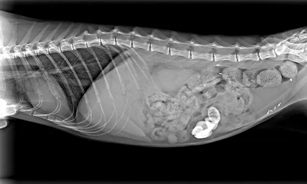
1. What do the radiographs (Figure 1) indicate?2. What would be the most likely diagnostic differentials?3. What is the next step?
Figures 1. Lateral (A) and ventrodorsal (B) full-body radiographs
Diagnosis
Enterolithiasis
Treatment & Outcome
Exploratory laparotomy was performed and foreign material removed from the ileocecocolic junction via a single enterotomy. The removed material appeared to be hair and other organic matter surrounded by a mineral shell (Figure 2). Based on the material’s gross appearance, the cat was diagnosed with enterolithiasis. Recovery was uneventful and, after a brief hospital stay, the patient was returned to the shelter for adoption.
Did You Answer?
1. The radiographs show several mineral-dense, opaque foreign bodies within the small intestine. Although an obvious obstructive pattern is not present, the size, shape, and number of objects present the opportunity for at least partial obstruction of the small intestine.2. The most likely diagnostic differential would include intestinal foreign bodies that would create a mineral-dense image on radiographs. Ingested items such as rocks are less common in cats than in dogs (in the author’s experience). Summation of nonmineral-dense materials would be possible but unlikely in such a successive pattern. Endogenously produced mineral opacities (eg, enteroliths) would be another possibility. 3. Although other imaging modalities (ie, ultrasound) could be considered, exploratory laparotomy was both diagnostic and therapeutic.
A Comment on Enterolithiasis
Although enteroliths are considered relatively common in horses, they are rare in dogs and cats. A review of the literature revealed only one report of enterolithiasis in a dog and one in a cat.1,2 In the latter, the inciting cause was speculated (but not confirmed) to be surgery to resolve pyometra 1 year earlier. In the present case, the medical and surgical histories were unknown, although the cat had been spayed—as evident from the apparent surgical material on radiography and confirmed during exploratory laparotomy.
KIRK MILLER, DVM, DABVP, is clinical instructor at Oregon State University where he teaches the small animal primary care rotation. He researches the natural incidence of heartworm disease in dogs in the Willamette Valley of Oregon and the safety and efficacy of a new feline spay technique. He completed his internship in small animal medicine and surgery at Angell Memorial Animal Hospital and earned his DVM from Colorado State University.