Perineal Herniation in a Bichon Frise
Sugar, an 8-year-old, 14-lb, intact male bichon frise, was presented with constipation and swelling under the tail.
History
Sugar was initially presented to his primary veterinarian for a 3-week history of tenesmus. Recommendations included adding psyllium fiber (metamucil.com) to Sugar’s diet and providing lactulose as a stool softener. Sugar’s owners recently noted swelling under the tail and, in the past 3 days, an inability to produce stool.
Related Article: Perineal Hernias
Examination
On referral, Sugar had a heart rate of 132 bpm, respiratory rate of 32 breaths/min, and temperature of 100.2°F. His mucous membranes were pink but tacky with a CRT of less than 2 seconds. He was approximately 5% dehydrated.
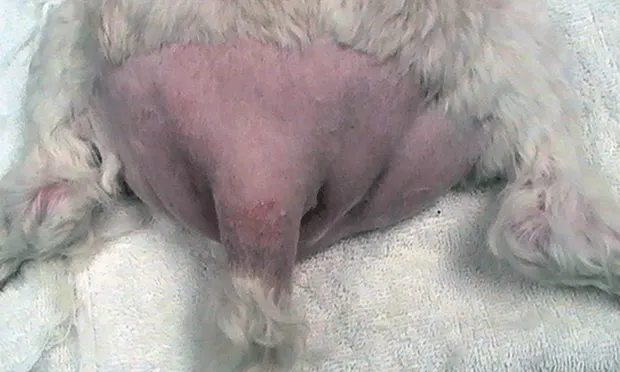
Although Sugar was overweight (BCS, 7/9), a distinct bulging was evident surrounding the perineum from the 3-o’clock to 9-o’clock positions (Figure 1). The swelling was soft, fluctuant, and reducible on gentle, external palpation on the right side but firmer on the left.
On rectal examination, using digital pressure in the lateral direction, lack of support was noted on the left and right sides of the rectal wall. The rectum was dilated and enlarged with bilateral outpouchings. Fecal impaction was noted on the left side. A large-yet-soft, symmetric prostate could be palpated on the cranial midline.
Figure 1. Distinct bulging was apparent ventral (1A) and lateral (1B) to the rectum.

Laboratory Diagnostics
PCV was 58% (range, 35%–55%), and total protein was 7.9 g/dL (range, 5.2–8.2 g/dL). No significant irregularities were noted in the serum biochemistry profile or CBC.
The author extends gratitude to Zachary Ricker, DVM, for providing photographs and Mark Rochat, DVM, MS, DACVS, for providing radiographs presented in this article.
Ask Yourself
Based on history and examination, what is the best next step for Sugar?
A. Perform castration because this condition should respond to elimination of testosterone alone.
B. Continue with conservative therapy, adding cisapride.
C. Recommend bilateral herniorrhaphies, colopexy, and castration.
D. Determine and eliminate the underlying cause (eg, prostatitis, cystitis, diarrhea, constipation, anal sacculitis), after which the condition should resolve.
E. Recommend a rectal pull-through to eliminate the diseased rectum.
Correct Answer

C. Recommend bilateral herniorrhaphies, colopexy, and castration.
Middle-aged, intact male dogs represent 90% of perineal herniation cases. The most commonly affected breeds include the Boston terrier, Pekingese, boxer, collie, and Old English sheepdog. Female dogs are rarely affected, possibly because of their larger levator ani muscle (used in parturition).
Atrophy in the pelvic diaphragm muscles seems neurogenic in origin. This atrophy results in lack of support for the rectum and thus a buildup of feces in the resulting redundant dilations. Herniation most commonly occurs between the levator ani and external anal sphincter.
Figure 2. Caudal aspect of lateral abdomen reveals marked dilation, sacculation, and redundancy of rectal tissue at the region of herniation.

Causes
The exact cause of perineal herniation is unknown, although many theories exist. A hormonal influence would be a logical assumption given the propensity for intact male dogs to have this problem.
The hormonal influence of the prostate via relaxin and the testicles via estrogen production may promote breakdown of the pelvic diaphragm. Although it is debatable, castration at the time of hernia repair may help prevent future herniation.
Approximately 10% to 51% of dogs with perineal herniation have true prostatic disease often manifested as prostatomegaly.1 Although a direct connection has not been made, an enlarged prostate may promote straining and thus breakdown of the pelvic diaphragm. Alternatively, these could be concurrent but unrelated age-associated conditions in older intact male dogs.
Figure 3. Primary closure of the muscle defect by apposing the levator ani and external anal sphincter.

Diagnosis
Diagnosis of perineal herniation is based on the clinical findings of perineal swelling (often reducible via external compression), and colonic dilation and lateral deviation (Figure 2.) resulting from loss of muscle support (confirmed by digital rectal examination). Unilateral cases provide asymmetry for comparison.
Clinical signs are related directly to herniated structures. The rectum is commonly dilated because of lack of support, leading to constipation and obstipation. The bladder, prostate, fat, and, less commonly, small intestines may be herniated. Bladder herniation must be addressed on an emergent basis and can be confirmed via radiography (± contrast cystography) or ultrasonography; it can also be verified (and relieved) with cystocentesis. Ultrasonography is warranted to rule out significant prostatic disease.
Figure 4. Elevation of the internal obturator flap from the ischial table.

Treatment
Surgical correction of perineal herniation is standard. The defect may be closed primarily by apposing separated muscles (Figure 3.). Because this is often insufficient, the most common method of repair is muscle transposition using the internal obturator as a muscle flap (Figure 4.). To close the herniation, the internal obturator muscle is elevated off the floor of the ischium, rotated, and then secured medially (often to the external anal sphincter), dorsally, and laterally (Figure 5.).
If the internal obturator is insufficient for repair, other options include superficial gluteal transposition, semitendinosus muscle transposition, fascia lata grafting, or the use of biomaterials (eg, porcine small intestinal submucosa, polypropylene mesh).
Figure 5. Internal obturator flap secured in place, reinforcing the closed defect.

Many surgeons recommend colopexy and cystopexy (or vas deferens pexy) for complicated cases or to prevent recurrence. These procedures keep the colon and bladder, respectively, tethered cranially so future herniation is avoided in the event of recurrence or contralateral muscle breakdown. Castration is recommended at the time of initial herniorrhaphy. Bilateral hernia repairs may be staged to avoid fecal incontinence that may result from bilateral damage to the caudal rectal nerves. Alternatively, colopexy and cystopexy may be performed first and herniorrhaphy 7 to 10 days later when swelling and inflammation have diminished.
Postoperative complications include incisional infection, temporary or permanent fecal incontinence (a concern with single-session bilateral hernia correction), sciatic nerve injury, tenesmus, recurrence (0%–70%),2 and urinary dysfunction.
Sugar underwent bilateral internal obturator muscle transpositions to repair his bilateral perineal hernias. Under the same anesthesia, colopexy and vas deferens pexy were performed before the herniorrhaphy procedure. Castration was also performed. Epidural bupivacaine was administered for increased peri- and postoperative comfort and to aid in disimpaction.
Outcome
Sugar was discharged from the hospital 48 hours postoperatively on tramadol (2.2 mg/kg PO q6–12h) and amoxicillin–clavulanic acid (13.75 mg/kg PO q12h for 10 days). A high-fiber prescription diet was initiated (Prescription Diet w/d Canine). Eight weeks later, the hernia repairs were intact and normal defecation had resumed. Rare episodes of straining were diminishing.
The Take-Home
Intact male dogs, especially the Boston terrier, Pekingese, boxer, collie, and Old English sheepdog, are most commonly affected by perineal herniation.
Perineal herniation is diagnosed by perineal palpation and rectal examination.
Imaging modalities (eg, ultrasonography) may clarify which structures are herniated as well as illuminate concurrent or underlying disease (eg, prostatic disease).
Although no direct connection has been documented, castration is recommended to eliminate any hormonal influence and prostatic involvement.
Internal obturator muscle transposition is the most common repair technique used for herniorrhaphy.
Colopexy and cystopexy may help prevent recurrence or contralateral herniation.
KRISTY BROADDUS, DVM, MS, DACVS, is small animal surgeon at Veterinary Emergency & Specialty Center in Richmond (Carytown) and Midlothian, Virginia. Her professional interests include orthopedic and soft tissue surgery. In addition to spending 3 years as an Oklahoma State University faculty member, Dr. Broaddus completed a rotating internship in small animal surgery and medicine and 3-year residency in small animal surgery at Auburn University, where she earned her master’s degree. Dr. Broaddus earned her DVM from Michigan State University.