Palpating for the Ortolani Sign When Diagnosing Hip Dysplasia
The biggest risk factor for the development of osteoarthritis of the canine hip joint is hip laxity. Laxity of the hips in young dogs can be detected by a clinical test, the Ortolani sign. The test was reportedly first described in 1938 by Marino Ortolani, who was professor and chief of pediatrics in Ferrara, Italy. The test is used to detect congenital luxation of the hip in newborn children and has been adopted by the veterinary profession to detect hip laxity in puppies. Since puppies develop hip laxity in the first few months of life, the test can be used beginning at approximately 4 months of age.
Step by StepPerforming the Ortolani Test
The Ortolani test is generally used in young dogs (4 to 12 months of age). The test is typically performed with the dog sedated or anesthetized because it can cause pain in dogs with hip laxity. With the patient in lateral recumbency, the test is performed in the following manner:
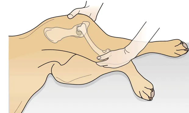
Put the hip at approximately a normal weight-bearing angle. Apply a dorsal force to the stifle joint, which, in a lax hip, will displace the femoral head dorsally beyond the dorsal acetabular rim. In a hip without laxity, the femoral head will not displace.While still applying this force, slowly abduct the limb and feel for the point at which the femoral head clicks, "clunks," or grates as it reduces back into the acetabulum-a positive result. If this sensation is not felt, the result is considered negative. Note the angle at which the hip reduces-the Ortolani angle.
When the patient is in dorsal recumbency, the preceding steps are followed, with modifications as shown in the photograph (note the angle of reduction).
Interpreting the Test Result1. A positive Ortolani test result indicates a lax hip. It is known that hip laxity is the biggest risk factor for development of osteoarthritis of the hip in dogs.1 The greater this hip laxity, the greater the probability that osteoarthritis will develop in this hip. However, this varies according to breed; some breeds (eg, rottweiler) are relatively more tolerant of laxity than others (eg, German shepherd).
2. In a clinical setting, if the clinician suspects that a young dog may have clinical hip dysplasia, the Ortolani test should be part of the examination and diagnostic protocol. Young dogs with hip dysplasia may be reluctant to exercise, may sit down when at exercise, may have lameness and joint stiffness, and will have pain on manipulation of the hip (full extension and often on abduction as well). If the history and clinical examination suggest hip dysplasia, the clinician should consider radiography and Ortolani testing.
Hips with No Visible Sign of DysplasiaTypically, radiographs of the hips are obtained in the ventrodorsal hip-extended position. However, although this projection is useful for determining symmetry and allows the clinician to evaluate the bony conformation and status of the hip joints, it underestimates hip laxity. This underestimation occurs because, when the hip joints are extended, the joint capsule is tightened; tightening tends to reduce subluxation of the femoral head. Other radiographic protocols, such as the PennHIP scheme, are designed to specifically measure passive hip laxity.
Severe Hip DysplasiaSince sedation is often required for good-quality pelvic radiographs in young dogs, the clinician has the opportunity to perform the Ortolani maneuver to test for hip laxity following radiographic evaluation. Hips such as the one shown above (8-month-old dog with positive Ortolani test result) are likely to have palpable laxity and a positive Ortolani test result (see Warnings). However, the Ortolani test should also be performed on hips with no visible sign of dysplasia as the hip-extended view may mask laxity that can be detected by physical examination.
SummaryIn young dogs, the Ortolani test is very useful for detecting hip laxity. The clinician needs to remember the limitations of the test but should also note the value of the test as an addition to other clinical and radiographic methods to evaluate hips.
Figures 1 & 2. Reprinted from British Small Animal Veterinary Association's Manual of Musculoskeletal Disorders. Houlton JEF, Cook JL, Innes JF, Langley-Hobbs SL (eds), with permission.
Procedure PearlWhen performing the test, note two things:• The angle of reduction. The greater it is, the greater the hip laxity.• The sensation when the femoral head reduces. This can provide the examiner with information on the integrity of the dorsal acetabular rim and the cartilage of the femoral head. A grating or crepitant sensation upon hip reduction suggests erosion and damage to the rim or femoral head.
Warnings• The Ortolani test is primarily used in dogs 4 to 12 months of age. Before 16 weeks of age, false-negative results are common.2• The development of osteoarthritis of the hip can cause false-negative results (ie, a negative Ortolani test result but a dysplastic and arthritic hip). This is presumably because the secondary changes to the joint, such as periarticular fibrosis, reduce palpable laxity, and the Ortolani test result becomes negative.3 Thus, in dogs over 1 year of age, the value of the Ortolani test is diminished-secondary changes have had time to occur.
PALPATING FOR THE ORTOLANI SIGN WHEN DIAGNOSING HIP DYSPLASIA • John Innes
References1. Advances in diagnosing canine hip dysplasia. Smith GK. JAVMA 210:1451-7, 1997.
Early detection of canine hip dysplasia: Comparison of two palpation and five radiographic methods. Adams WM, Dueland RT, Meinen J, et al. J Am Anim Hosp Assoc 34:339-347, 1998.3. Relationships between results of the Ortolani method of hip joint palpation and distraction index, Norberg angle, and hip score in dogs. Puerto DA, Smith GK, Gregor T, et al. JAVMA 214:497-501, 1999.