Orthopedic Examination of the Forelimb in the Dog
Derek B. Fox, DVM, PhD, Diplomate ACVS, University of Missouri–Columbia
Dogs bear most of their weight with the thoracic limbs. As a result, orthopedic disease of the forelimb is particularly challenging. For examination purposes, the forelimb can be anatomically divided into the paw, antebrachium, brachium, scapula, and interpositioned joints. Causes of lameness, and the anatomic locations they effect, will differ by the age, breed, and lifestyle of the dog. Thus, the orthopedic examination can be tailored to focus more on areas prone to disease based on patient signalment. However, all anatomical aspects of the limb should be assessed in every patient.
Orthopedic examination of the forelimb starts with observing the animal ambulate at various speeds. Weight-bearing lameness of the front limb typically results in a "head bob" in which the head is lifted during weight-bearing of the painful limb. Animals affected bilaterally may not exhibit an obvious head bob but can show a shortened stride of each limb. A full neurologic examination should accompany the orthopedic examination to rule out neurologic causes for pain or lameness, such as a nerve root signature sign secondary to a laterally herniated intervertebral disk or brachial plexus pathology.
Step-by-Step: Orthopedic Examination of the Forelimb
Step 1: The Paw
Examination of the paw generally consists of assessing each digit, including the nail and nail bed (A). Common sources of lameness arising from these sites include split nails, nail-bed tumors, fracture/luxation of the phalanges, penetrating foreign bodies, and pad lacerations. Careful palpation of the digits and manipulation of the joints in flexion and extension (B, C), while observing for evidence of pain or swelling, make up a fairly sensitive method to determine whether any part of the paw is a source of pain. Injury to the paw or digits is a frequent source of a non-weight-bearing lameness. Because many dogs are sensitive to having their paws handled, the examiner should also palpate the feet from the unaffected legs to gain a sense of the level of an animal's reluctance to having its paws manipulated.
Procedure Pearl
Careful palpation of the digits and manipulation of the joints in flexion and extension make up a fairly sensitive method to determine whether the paw is painful.

FIGURE A
Step 2: The Carpus
Various conditions affecting the carpus include degenerative joint disease, suppurative arthritis, subluxations or luxations from traumatic hyperextension, fractures, and carpal laxity syndrome in young dogs. Nontraumatic joint inflammation of the radial carpal joint is fairly easy to feel as a depressible distension of the joint capsule (A). Palpation of the carpus should include placing the joint under stress in extension, flexion, abduction, and adduction for the observation of excessive mobility or pain in any of these planes. The carpus should have minimal abduction and adduction, may extend 5º to 10º (B), and should be able to be flexed comfortably until the palmar surface of the paw nearly touches the flexor surface of the antebrachium (C).

FIGURE A
Step 3: The Antebrachium
Sources of lameness that can arise from the radius and ulna in the young dog include hypertrophic osteodystrophy, eosinophilic panosteitis, and physeal disturbances; and in young adult and adult dogs fractures, neoplasia, angular limb deformities, and hypertrophic osteopathy. Examination of the antebrachium should focus on deep palpation of both bones in an attempt to elicit an osseous source of pain or to detect focal areas of swelling. The relationship of the carpus to the elbow should also be observed as evidence of angulation from physeal damage and abnormal development of the radius and ulna. Aberrant growth from physeal disturbances can result in varus, valgus, procurvatum, recurvatum, or rotational or translational deformities.
Step 4: The Elbow
The elbow is primarily a hinge joint that optimizes flexion and extension but allows limited abduction and adduction and only slight rotation. Orthopedic diseases affecting the elbow include fragmented coronoid disease, ununited anconeal process, osteochondritis dissecans, various fractures, congenital luxations, and radioulnar incongruity in the young dog and degenerative joint disease, suppurative arthropathies, fractures, subluxations, luxations, and neoplastic processes in the adult dog. Examination of the elbow consists of putting the joint through its range of motion in flexion (A) and extension (B) while feeling for the presence of crepitus and observing for a painful response. Hyperextension should be accompanied by only minimal or no discomfort in a normal dog. While the distal humerus is held, the antebrachium is abducted and adducted in an attempt to elicit pain or excessive motion consistent with collateral ligament damage and subsequent joint laxity. Elbow joint effusion can be palpated for by digitally feeling both distal humeral epicondyles simultaneously with index finger and thumb, while drawing fingers caudally toward the olecranon (C). Normally a concave depression exists between these structures. With effusion, however, this area can become distended. With the chronicity of the abnormality come hypertrophy and thickening of the joint capsule, which will palpate more firmly. Specific pain localization may be detected with fragmented coronoid disease by applying digital pressure over the medial coronoid and looking for a painful response.
Procedure Pearl
A shoulder abduction tests the integrity of the medial joint capsule and collateral ligament.

FIGURE A
Step 5: The Brachium
Conditions potentially affecting the canine brachium include panosteitis, hypertrophic osteodystrophy, and fractures in young dogs and fractures, neoplasia, and hypertrophic osteopathy in the adult dog. One of the most common tumors of the canine forelimb is osteosarcoma, which more frequently forms in the proximal humerus and distal radius and ulna. Examination of the brachium essentially consists of deep palpation along the length of the bone to examine for evidence of pain and areas of inflammation or swelling.
Procedure Pearl
Examination of the brachium consists of deep palpation to examine for evidence of pain and inflammation.
Step 6: The Shoulder
The shoulder joint is considered a ball-and-socket joint that possesses both passive and active stabilizers. Normal motion includes extension and flexion, abduction and adduction, and internal and external rotation. Passive stabilization of the joint is provided by both medial and lateral glenohumeral ligaments, the shape of the humeral head and glenoid, and the cohesiveness of joint fluid. Active stabilization of the shoulder is maintained by the musculotendinous units of the rotator cuff: the supraspinatus, infraspinatus, teres minor, and subscapularis. As with orthopedic conditions of the dog involving other structures, sources of pain can depend on the age of the animal.
Young dogs with shoulder pain should be examined closely for osteochondritis dissecans, whereas in the adult dog common sources of lameness localized to the shoulder can include degenerative joint disease, biceps tenosynovitis, shoulder instability or luxations, mineralization of the supraspinatus, infraspinatus contracture, ununited caudal accessory process of the glenoid, articular fractures, and other causes of suppurative arthropathies.
Procedure Pearl
A large component of shoulder joint examination is putting the joint through a full range of motion to observe for excessive excursion or elicitation of pain.
A large component of the examination of the shoulder joint is putting the joint through a full range of motion to observe for excessive excursion, or elicitation of pain in any particular direction. For example, the joint should be comfortably flexed (A) and extended (B), internally and externally rotated, and abducted and adducted.
Limitation in internal rotation can prompt suspicion of contraction of the infraspinatus tendon.
A shoulder abduction can also be completed to test the integrity of the medial joint capsule and collateral ligament, during which the angle between the scapula and humerus is measured via goniometry during abduction. Abduction greater than approximately 32º can lead to suspicion of medial shoulder instability (C). A shoulder drawer examination, in which the proximal humerus is translated cranially and caudally with respect to the glenoid, can be completed to assess for instability of the joint in either direction (D). The biceps tendon should be palpated as it originates on the supraglenoid tuberosity and courses down the intertubercular groove for evidence of inflammation (E). Deep digital pressure is applied to the biceps tendon while flexing the shoulder and extending the elbow in an attempt to elicit pain as supportive evidence for biceps tenosynovitis (F).
Procedure Pearl
Orthopedic examinations should be followed up with radiography, ultrasonography, or other imaging to confirm diagnoses.

FIGURE A
Step 7: The Scapula
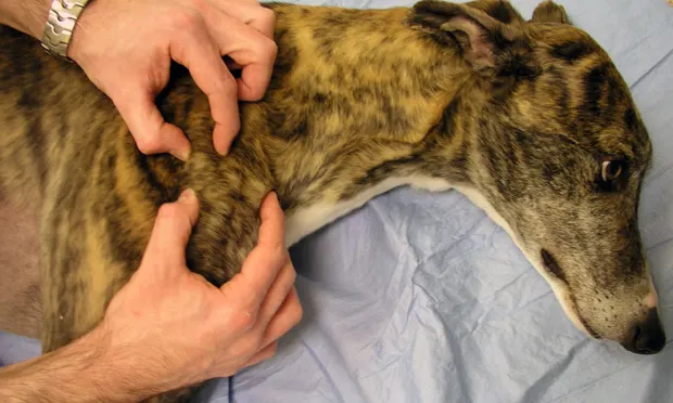
Although the scapula is the least frequently involved structure causing canine forelimb pain, certain conditions, such as tumors and fractures, can affect it. Furthermore, the supraspinatus and infraspinatus muscles and prominence of the interposed acromial spine should be felt for muscle atrophy as evidence of any musculoskeletal source of forelimb disuse or many neurologic conditions. The muscular attachment of the scapula can also be avulsed from the thoracic wall with certain types of trauma. Scapular avulsion is primarily diagnosed by palpation of excessive excursion of the scapula from the chest wall since radiographs can look deceptively normal.
Procedure Pearl
Scapular avulsion is primarily diagnosed by palpation since radiographs can look deceptively normal.
Conclusion
Observation of gait, neurologic examination, and systematic palpation of specific anatomic structures will allow the elucidation of a majority of orthopedic conditions affecting the canine forelimb. Most often, presumptive diagnoses based on orthopedic examination should be followed up with radiography, ultrasonography, or other imaging to confirm the diagnosis and provide clients with treatment options and prognoses.
Acknowledgment: The author thanks Dr. David Dismukes for his assistance in preparing the figures.