Oral Cavity Disease in Cats
Andrew Sparkes, BVetMed, PhD, DECVIM, MANZCVS, MRCVS, Simply Feline Veterinary Consultancy, Dorset, United Kingdom
In the Literature
Falcão F, Faísca P, Viegas I, de Oliveira JT, Requicha JF. Feline oral cavity lesions diagnosed by histopathology: a 6-year retrospective study in Portugal. J Feline Med Surg. 2020. doi: 10.1177/1098612X19900033
The Research …
A substantial retrospective study evaluated 297 surgically removed feline oral cavity lesions submitted to a pathology laboratory service in Lisbon, Portugal, over a 6-year period. Most samples were incisional (86.2%) or punch biopsies (1.7%), and 12.1% were excisional biopsies.
Overall, 64.4% of biopsies were from cats ≥7 years of age and 31.9% were from cats 7 months to 6 years of age. The major anatomical regions biopsied were the gingiva (43.1%), oral mucosa (16.2%), tongue (10.8%), lips (8.1%), and palate (5.7%). Other sites included the oropharynx, maxillary bone, salivary glands, floor of the mouth, and tonsils. Histopathology revealed an inflammatory process in 63% of cases and a neoplastic process in 37%. The proportion of neoplastic cases increased with age and accounted for most lesions in cats ≥11 years of age. However, even in young cats (ie, 7 months to 2 years of age) and young adult cats (ie, 3-6 years of age), neoplastic lesions were seen frequently, representing 5.4% and 16.7% of biopsies in cats of those ages, respectively.
Of the 187 cases of inflammatory disease, 62% (n = 116) were feline chronic gingivostomatitis (FCGS), with the most common sites affected being the gingiva (n = 66), oral mucosa (n = 22), and oropharynx (n = 12). FCGS was seen in cats of all ages but was most common in male cats 7 months to 10 years of age. Eosinophilic granuloma complex (EGC) lesions accounted for 17.6% (n = 33) of inflammatory lesions and comprised 23 eosinophilic ulcers and 10 eosinophilic granulomas. The most common sites of EGC lesions were the lips (n = 12) and tongue (n = 5). Other inflammatory lesions included nasopharyngeal polyps, nonspecific stomatitis, and gingival hyperplasia.
Of the 110 neoplastic lesions, 90 (81.8%) were classified as malignant, and overall, squamous cell carcinoma (SCC) was the most common tumor (n = 49; 44.5%). The gingiva (n = 18) and mandible (n = 10) were the most common sites affected by SCC, and most of these cats were ≥11 years of age. Other neoplastic lesions included undifferentiated tumors (17.3%), odontogenic tumors (8.2%), peripheral nerve sheath tumors (8.2%), adenocarcinomas (5.4%), and fibrosarcomas (4.5%).
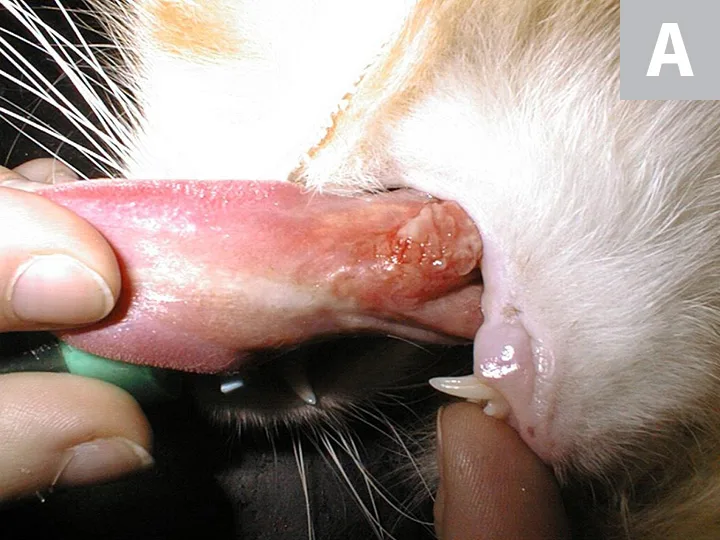

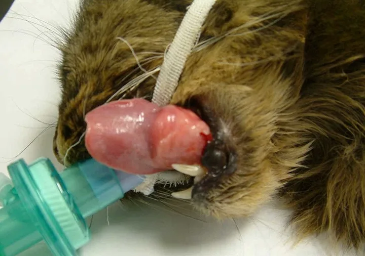
Two cats with sublingual mass lesions in which the importance of biopsy and histology is emphasized. The lesion in Figure A was diagnosed as an eosinophilic granuloma complex legion and the lesion in Figure B as squamous cell carcinoma.
… The Takeaways
Key pearls to put into practice:
This study emphasizes the breadth of pathologies that occur in the feline oral cavity and the need for histologic examination of biopsy material for an accurate diagnosis.
In this study, inflammatory lesions were more common than neoplastic lesions, with FCGS and EGC being the most common. This emphasizes the importance of differentiating the disease process, as underlying etiologies and management options differ markedly.1
Although neoplastic disease is more common in older cats, oral tumors can also be seen in younger cats, and the presence of malignant disease does not invariably equate to a poor prognosis. Dependent on the underlying disease, different treatment options may be possible.2-4
You are reading 2-Minute Takeaways, a research summary resource presented by Clinician’s Brief. Clinician’s Brief does not conduct primary research.