Ophthalmic Screening for Systemic Hypertension in Cats
Ronald Spatola, DVM, MS, DACVO, The Animal Eye Institute, Cincinnati, Ohio
In the Literature
Moretto L, Lavaud A, Suter A, Günther C, Pot S, Glaus T. Reliability of detecting fundus abnormalities associated with systemic hypertension in cats assessed by veterinarians with and without ophthalmology specialty training. J Feline Med Surg. 2021;23(10):921-927.
The Research ...
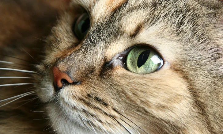
Fundoscopy is essential in screening for ocular and systemic disease in cats and allows quick, noninvasive, direct visualization of neurologic and vascular tissue. Detection of fundic lesions can lead to early recognition of disease processes, helping direct further diagnostics and treatment.
This study compared the ability of a recent veterinary graduate and a board-certified veterinary ophthalmologist (or veterinary ophthalmology resident under direct supervision) to detect retinal lesions associated with systemic hypertension (SHT) in cats. Cats were client-owned and had suspected hypertensive target organ damage (TOD) or were otherwise at risk for SHT with initial elevated systolic blood pressure (BP). Cats with TOD and BP >160 mm Hg were considered truly hypertensive, and cats without TOD with serial BP measurements <160 mm Hg were controls. The recent graduate used indirect ophthalmoscopy with a 28-diopter condensing lens; the ophthalmologist used 28-, 20-, and/or 15-diopter lenses.
Of 33 cats, 27 were confirmed to have SHT based on TOD or subsequent BP measurements. Veterinary ophthalmologists were able to detect fundic lesions in 24 cats with SHT. The recent graduate was able to detect fundic lesions in 19 of the 24 cats (72% sensitivity), as well as fundic lesions (including hemorrhage and retinal detachment) in every cat presented for blindness, but subtle lesions (eg, focal retinal edema, petechial retinal hemorrhages, peripheral retinal vascular tortuosity) were more likely to be missed. A training effect was observed; the recent graduate was less likely to miss lesions as the study progressed.
Results suggest nonspecialty-trained clinicians can detect routine fundic lesions in cats with SHT, especially with practice; however, detection of subtle lesions may require evaluation by a clinician trained in veterinary ophthalmology.
... The Takeaways
Key pearls to put into practice:
Indirect ophthalmoscopy can help screen for ocular and systemic disease, especially in patients with systemic hypertension and/or vision loss.
Regularly practicing fundoscopy can help sharpen skills and lead to more accurate detection of fundic lesions. A Finoff transilluminator and condensing lens (28 diopter or 2.2 pan retinal) is sufficient for clinicians without specialty ophthalmology training.
Patients with suspected systemic and/or ocular disease with no evidence of ocular lesions detected by the primary clinician should be referred to a veterinary ophthalmologist to identify subtle lesions and avoid a delay in diagnosis and treatment.
You are reading 2-Minute Takeaways, a research summary resource presented by Clinician’s Brief. Clinician’s Brief does not conduct primary research.