Occlusal Issues
Judy Rochette, DVM, FAVD, DAVDC, West Coast Veterinary Dental Services
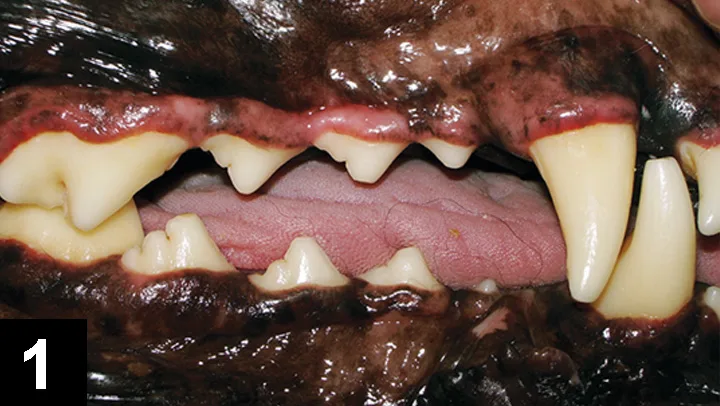
Normal canine occlusion
You have asked...
What are the most common occlusal issues in dogs and cats, and how are they treated?
The expert says…
Before any discussion about malocclusions (ie, misaligned teeth) can begin, a thorough understanding of healthy occlusions is necessary. The wild phenotype of domestic dogs and cats has interdigitating cheek teeth that create a pinking shears effect (Figures 1 and 2), a mandibular canine tooth that interdigitates with the maxillary third incisor and canine tooth, and mandibular incisors that rest on the cingulae of the maxillary incisors. This model of occlusion has been naturally selected as the most efficient and durable for grabbing, holding, tearing, and masticating a predator’s diet.

Normal feline occlusion
The canine and feline facial architecture complements this oral arrangement with stress- and/or load-bearing reinforcement of the facial bone, where maximum forces occur. Historically, humans have created new phenotypes for dogs and cats and selected for genes that would normally be selected against in nature. What is considered acceptable as a breed standard may not be ideal for quality of life.
Malocclusions may result in self-induced soft-tissue trauma or repetitive concussive dental trauma. These can cause irreversible pulpitis, dysmastication, and tooth crowding, which may make an area prone to periodontal disease.
Malocclusions, or misaligned teeth, may result in self-induced soft-tissue trauma or repetitive concussive dental trauma.
Malocclusions can be dental (ie, jaw lengths are correct but one or more teeth are displaced or improperly oriented) or skeletal (ie, discrepancy between jaw lengths is responsible for the occlusal issues).
Dental Malocclusions (Class 1)
Class 1 dental malocclusion may be assigned to a tooth that is tipped, rotated, and/or displaced relative to its expected position and orientation within the dental arcade.
Some dental malocclusions may be transmitted genetically. Mesioversion of maxillary canine teeth has a strong familial tendency in the Shetland sheepdog, but this condition is now seen in other canine and feline breeds. Palatoversion of mandibular canine teeth also has a strong genetic heritage, as orientation of the tooth buds and retention of deciduous canine teeth are both genetically controlled. Some dental malocclusions are truly a developmental accident or trauma induced, resulting in phenotypic growth that does not represent the animal’s genotype; these exceptions are the minority.
Contact of the Maxillary Fourth Premolar with Buccal Mandibular Molar Tissues in Cats
This class 1 occlusal problem, which does not have an obvious instigating cause, can be found in cats of any breed or head type. When the tip of the major cusp of the maxillary fourth premolar contacts the soft tissues on the vestibular aspect of the mandibular molar tooth, the insult can range from the soft tissue ulceration and pain to bone loss, which can compromise the periodontal health of the mandibular molar (Figure 3). Signs may include head shaking, pawing at the affected side, and/or sudden cessation of or aversion to eating. If untreated, the chronic soft tissue irritation can lead to formation of hyperplastic granulation tissue, which can physically mimic a soft tissue tumor.

Soft tissue damage buccal to the mandibular molar from the maxillary fourth premolar in a cat.
Treatment involves extraction of the offending tooth (ie, maxillary fourth premolar). On occasion, damage to periodontal tissues may be so severe that the mandibular molar is compromised and should also be extracted. Rarely will mandibular molar extraction alone be sufficient. Caution must be exercised with any attempt at blunting the cusp tip, as the enamel and dentinal layers are thin. Blunting can easily lead to pain by exposing dentinal tubules or even pulp and subsequent loss of vitality. Given the extreme difficulty in treating this tooth endodontically, the end result is almost always extraction, hence the recommendation to extract as the first line of therapy.
Mesioversion, Palatoversion, & Linguoversion
Mesioversion refers to any tooth that is in its correct anatomic position but abnormally angled mesially or toward the midline at the front of the face (Figure 4). Palatoversion refers to a tooth that is tipped toward the palate. Linguoversion describes a tooth that is tipped toward the tongue.

Mesioversion of the maxillary canine tooth in a dog
Mesioversion of maxillary canine teeth can impede the mandibular canine teeth from occluding in their proper positions, resulting in buccal tipping and tooth-on-tooth contact, which can lead to concussive pulpitis and pain. The misoriented maxillary canine tooth can cause crowding and possible subsequent periodontal disease of the adjacent maxillary incisor. The affected canine tooth may be poorly erupted with pericoronitis or, in severe cases, unerupted with possible dentigerous cyst formation.
Teeth in mesioversion, palatoversion, or linguoversion can prevent the mouth from closing, creating an open bite. Palatoversion of the maxillary canine teeth (Figure 5) can cause extensive trauma to the mouth floor and/or the tongue. Linguoversion of the mandibular canine teeth can cause extensive palatal mucosal trauma with subsequent possible oronasal fistula formation, damage to the alveolar ridge with subsequent bone loss, or disruption of the periodontal apparatus supporting the maxillary canine tooth, with possible loss of that tooth to periodontal disease. Affected dogs are often head-shy, as any head handling may cause them to bite themselves; these dogs should not be placed in a head halter or collar until the bite has been corrected.

Feline palatoversion. Photo courtesy Dr. Bryan Hall
Mesioversion, palatoversion, and linguoversion can be treated by extraction of the offending canine tooth or with placement of an orthodontic appliance. Alternatively, the height of the crown can be reduced, but only if the practitioner can provide immediate endodontic treatment. Clipping, whether of a deciduous or permanent tooth, without immediate endodontic treatment will expose pulp chamber to infection and pain.
Examples of Class 1 Malocclusions
Mesioversion: Any tooth in correct anatomic position but abnormally angled mesially or toward the midline at the front of the face
Palatoversion: A tooth tipped toward the palate
Linguoversion: A tooth tipped toward the tongue
Skeletal Malocclusions (Class 2 & 3)
Skeletal malocclusions result from discordant growth of one or more areas of tooth-bearing bones. The more severe the skeletal malocclusion, the farther the facial contours are from the wild phenotype. Teeth may be correctly positioned within their discrete area of bone, but the overall occlusion is incorrect because the bone fails to mimic the ideal phenotype. The shape and length of the upper arcades are closely associated with the genes for head type, with the brachygnathic maxilla tied to the brachycephalic genotype. The final contours of each lower arcade are determined by two growth centers within each jaw. The first is located in the area of the ramus, a site that is completely under genetic control but, unlike the maxilla, is not tied to head type. Abnormal growth from this area can be detected by loss of interdigitation of the cheek teeth. Animals with these skeletal malocclusions should be culled from the breeding pool, as this bite will likely be passed to the offspring. A second growth center is located in the area of the lip fold, and influences on this center tend to be multifactorial. An animal with normal cheek-tooth interdigitation but one or more incisor teeth in an even, or reverse, scissor may not be as certain to have affected progeny. The degree of affectedness in either the maxilla or mandible can vary (eg, severity of maxillary brachygnathism and mandibular prognathism in bulldogs). Regardless, skeletal malocclusions have a genetic basis, and owners should be made aware of this when selecting breeding stock.
Jaw Length Discrepancy
When an abnormal rostral–caudal relationship between the dental arches exists, there is potential for soft tissue trauma, rotation of teeth, and crowding. These situations may predispose the area to periodontal disease development and loss of function. Selective extractions can reduce soft tissue damage and crowding. Occasionally, orthodontics can move individual teeth, or whole sections of an arch, into a more physiologic position. In cases of severe jaw length discrepancies, the teeth will never function as intended.
Closing Remarks
The ultimate goal is to offer the patient a pain-free, functional mouth that stays in a relative state of equilibrium and health. Veterinarians should strive to guide owners and breeders toward making treatment decisions that can sustain the long-term oral health.