Obtaining a Diagnostic Electrocardiogram
Kristie Garcia, LVT, VTS (Cardiology), Garden State Veterinary Specialists, Tinton Falls, New Jersey
Obtaining diagnostic electrocardiograms (ECGs) is an often overlooked but valuable skill veterinary nurses can perform in practice. An ECG is a graphic recording of electrical activity occurring in the heart. ECGs always provide some patient information, but a properly obtained ECG of diagnostic quality can help the veterinarian evaluate the size of the patient’s heart chambers and identify the source of any abnormal heartbeats, so it is essential a recording be correctly obtained.
Following is a step-by-step explanation for veterinary nurses to better understand how to obtain an ECG recording that allows accurate interpretation of the results. (See Requirements for Obtaining a Diagnostic ECG.)
Requirements for Obtaining a Diagnostic ECG
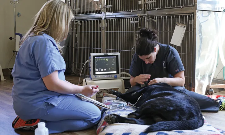
Place the patient in right lateral recumbency.
Place the limb electrodes on the patient as labeled; each electrode should be labeled with the limb to which it is to be attached. Color coding is optional.
The electrocardiogram is recorded for several seconds in each of the 6 limb leads, plus a longer recording in lead II at a known paper speed (50 mm/sec and 25 mm/sec are accepted in veterinary medicine) for rhythm evaluation.
Print out the electrocardiogram recording for the veterinarian, making sure to note the patient’s position.
Setting Up For an ECG
Have the following on hand when preparing to record an ECG:
ECG recording machine
Alcohol and/or ultrasound gel (See Figure 1.)
Alligator clips or ECG patches (See Figures 2 & 3.)
Blanket

FIGURE 1
Ultrasound gel and alcohol are used to facilitate conductivity between the leads and the patient.
Each of the electrodes are color coded and labeled with the correct limb where they will be placed on the patient: white/RA (right arm, or right front leg), black/LA (left arm, or left front leg), red/LL (left leg, or left rear leg). Many ECG recorders have a 4th electrode (green, attached to the right rear leg), which is a ground electrode. (See Figure 3, above.)
Patients should always be placed in right lateral recumbency to record a diagnostic ECG. This position is standard for correct ECG recording because the standardized measurements were all obtained in this position. Placing the patient in right lateral recumbency is sometimes unrealistic; in these cases, the ECG may still be valuable for assessing the heart rate and rhythm but is not a true diagnostic ECG.
Recording an ECG in another position is not wrong, but measurements of the waveforms cannot be taken on a recording made in any position other than right lateral recumbency, and it is important to note the different position in the patient record. This type of recording is typically of lead II and referred to as a rhythm strip.
Step 1
The patient should be placed in right lateral recumbency; if this is not an option, note the patient’s position in the chart and do not make mean electrical axis (MEA) or other calculations.
Electrodes should not touch each other or the metal table because this produces an artifact.
Tip
When placing electrodes:
Right side: Snow over grass (ie, white electrode front leg; green electrode rear leg)
Left side: Smoke over fire (ie, black electrode front leg; red electrode rear leg)
Right lateral recumbency: Snow and grass on the ground.
Step 2
Attach limb electrodes to the patient. Electrodes should be attached at the elbows and the stifles. Limbs should be held parallel to the floor and perpendicular to the long axis of the patient. (See Figure 4.)

Leads placed on a patient at the elbows and stifles, the correct positions for an ECG recording.
If the clips have alligator teeth, consider blunting or flattening the clips to make the patient more comfortable. If the leads are snapped on with adhesive patches, the area must be clipped and cleaned before the adhesive patches are applied.
A shaking or panting patient will produce an artifact on the recording. In this case, move the electrodes distal on the limbs away from the moving body wall. Spray alcohol on the electrodes where they contact the patient to ensure clear conductivity. Electrode or ultrasound gel is also acceptable and can be especially useful when a patient needs to be monitored for long periods (ie, surgery) because unlike alcohol, gel does not evaporate quickly.
Step 3
Record the electrocardiogram at 50 mm/sec (25 mm/sec is also acceptable in some patients) and 10 mm/1 mv, which is standard amplitude calibration.
The ECG analysis should be made in lead II, which should always be labeled, along with the paper speed and calibration. It must be clearly noted in the patient chart if a different lead is recorded. (See Figure 5.)

An ECG printout: Note the paper speed and amplitude setting.
Step 4
Interpret the findings in context with the patient’s clinical signs, physical examination findings, and other diagnostics.
6 Lead ECG Readings
Standard Limb Leads
Lead I: RA to LA
Lead II: RA to LL
Lead III: LA to LL
Augmented Unipolar Limb Leads
aVR: RA to average of LA and LL
aVL: LA to average of RA and LL
aVF: LL to average of RA and LA
Conclusion
Knowing how to obtain an accurate electrocardiogram is a necessary skill for veterinary nurses as part of their charge to provide patients with the highest possible level of care. A properly obtained, correctly labeled electrocardiogram can help the attending veterinarian determine or confirm a diagnosis for the patient.