Nonhealing Wounds
Bryden J. Stanley, BVMS, MACVSc, MVetSc, DACVS, Michigan State University
In most cases, wounded animals heal quickly and effectively. This is partly because skin laxity in dogs and cats allows contraction to play a large role in healing; in addition, animals do not experience human comorbidities such as smoking and drinking. There are times, however, when veterinarians are faced with a wound that does not appear to be healing.
Levels of Care
Wound care practitioners must be familiar with normal wound healing so that any delay can be recognized. Many wounds perceived to be nonhealing are simply not provided with optimal conditions for healing. When faced with an atypical or nonhealing wound, the practitioner should scrutinize:
Wound management methods
Systemic influences affecting healing
Specific wound factors.

Wound Management
Tension is the enemy of wounds. Manipulating wound edges during suturing will indicate how to close with least resistance. If tension is too great for direct apposition, other techniques must be used (eg, tension sutures, relaxing incisions [Figure 1], skin stretching, or other forms of reconstruction). It is preferable to allow a wound to heal by second intention, rather than to close it under too much tension.

Figure 1. Wound closed under too much tension resulting in marked congestion due to venous compromise (A). Tension can be relieved with multiple, punctate incisions to allow expansion (shown here in a different wound) (B).
Prolonged pressure on skin over bony prominences will result in ischemia, necrosis, and the development of a decubital ulcer (Figure 2). These pressure sores result from inadequately padded bedding or poor bandaging. A variety of padded donuts, slings, beds, whirlpool, and physical nursing regimens have been described. There may be value in some topical wound stimulating medications such as tripeptide-copper complex or acemannan- or maltodextrin-based medications.
Figure 2. Formation of a decubital ulcer due to prolonged pressure over the greater trochanter following orthopedic repair. The full extent of ulceration was significantly larger than the cutaneous defect.

High motion areas such as the axilla, inguinal area, lip commissure, and joints are subject to repeated shearing forces that disrupt healing. This should be anticipated and definitive closure with strict immobilization is preferred until epidermal integrity is established. A combination of pressure and movement can impair the healing of footpad wounds. Special bandages, such as donut-type or clamshell bandages are needed, and relief from weight-bearing for 4 to 6 weeks may be necessary.
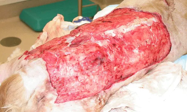
Wounds containing devitalized tissues have prolonged inflammation and delayed healing. Contaminated and dirty wounds must be meticulously debrided (often several times), both surgically and with intermittent lavage (Figure 3). Inadequate surgical debridement is a common reason for dehiscence.


Figure 3. Large amount of devitalized tissue following degloving injury (A).Wound appearance following surgical debridement (B).
Wounds in small animals are highly exudative in the early stages of healing. If a dressing is too occlusive or becomes wet, maceration of the wound will result. This delays healing and the wound will require daily lavage and an absorbent dressing for 24 to 48 hours.
Desiccation damages fibroblasts, epithelial cells, and capillary buds. Wound surfaces must be kept somewhat moist to promote epithelialization and protect fragile cells. The principle of moist wound healing is being used more in veterinary medicine. Moisture-retentive dressings can be applied to small animal wounds once exudate lessens.
Prevention of self-induced trauma should be routine. This can be accomplished with padded bandages, splints, side braces, Elizabethan collars (which may be reversed), wire sutures, and chemical restraint.
Systemic Influences
Malnutrition (less than 2 g/dl serum protein) can impede wound healing and interfere with the ability to fight infection. Supplemental nutrition, preferably enteral, should be instituted early in management. Efforts to normalize azotemia in uremic animals should be instituted
Corticosteroids impair early wound healing, slow down fibroplasias, and retard epithelialization. Cushingoid patients and those on high doses of corticosteroids respond better with a tension-free closure. Closure in several layers should be employed, and sutures should be left in longer (21 days). Epidermal integrity can be tested by removing a few sutures and trying to spread the incision apart. If the incision appears to be holding under this tension, the remaining sutures can be removed.
Elderly patients require meticulous nursing care, a high plane of nutrition, and attention to enhancing the blood supply to the wound. Early reconstruction, rather than second intention healing, is preferred. Vacuum-assisted closure devices may prove useful in these patients.
Specific Wound Factors & Diagnostics
If wound management issues have been addressed and systemic influences on wound healing ruled out, the nonhealing wound itself needs to be examined more closely. Neoplasia (benign or malignant) may be erosive rather than proliferative, and can appear similar to granulation tissue. Biopsy and histopathology will facilitate the diagnosis of neoplastic, fungal, pythiotic, immune-mediated, granulomatous, and other lesions.
Chronic infection seriously impacts wound healing. Macerated tissue culture is the most reliable way to determine the etiology of a serious wound infection, as superficial contaminants are not representative. The wound should be cleaned with sterile saline, then a biopsy taken aseptically from the deep, affected tissues. Aerobic, anaerobic, mycobacterial, and fungal cultures should be specifically requested.
Radiographic evaluation of nonhealing wounds should be routine. This may highlight a radiopaque foreign body or an underlying painful orthopedic issue, such as osteomyelitis, sequestrum, or arthritis. Ultrasonography is sometimes useful in wounds with deep extension-it may detect sinus tracts, foreign bodies, or underlying masses.
When to Consider Referring

Severe trauma cases exhibiting tissue devitalization respond best to debridement followed by early reconstruction to restore blood supply to the region (eg, robust skin flap, pedicled muscle flap, microvascular free tissue transfer) (Figure 5). Severe soft tissue injuries with exposed bone may also respond to wound-healing stimulants (acemannan-based medication) and forage, which provides a blood clot matrix over the exposed bone, encouraging granulation tissue formation.
Figure 5. Severe crush injury to the carpus with compromised blood supply (A). Reconstruction with a flexor carpi ulnaris muscle flap (B).
The true extent of a chronic decubital ulcer can be significantly larger than the visible defect. If a chronic ulcer fails to respond to good nursing care, infection control, and nutritional support, then referral for extensive debridement and reconstruction is indicated.
Successful closure of chronic axillary or inguinal wounds involves resection of skin edges and often a skin flap to reduce shear forces. Omentum will also enhance perfusion to the area. In addition to surgical techniques, immobilization of the axillary and inguinal areas is recommended. Cats have a tendency to form indolent axillary ulcers.
Radiation therapy will delay healing, whether administered before or after surgery. Wounds in irradiated tissue pose a huge challenge, and bringing blood supply in from outside the irradiated zone (eg, axial pattern flap, robust subdermal plexus flap, free vascularized graft) is recommended. There will be higher complication rates with these surgeries, and several revisional procedures may be required.
Infected wounds generally respond to aggressive open wound management and appropriate systemic antibiotic therapy, but unusual infections with such organisms as atypical mycobacteria, Nocardia, Actinomyces, Pythium, and fungal infections induce chronic nonhealing wounds. These infections can be difficult to definitively diagnose and are also a challenge to cure, with a combination of prolonged antibiotic therapy and wide excisional surgery necessary.
Whenever a draining tract is present, a foreign body should be suspected. The discharge can vary from serous to purulent, as secondary infection is usually present. These wounds should be biopsied, cultured, radiographed, and ultrasounded. The most useful diagnostic tool, however, is sinography, preferably under fluoroscopy or computed tomography (Figure 8). Due to their migratory nature, finding the foreign body can be a challenge and exploration should be thorough. Once the foreign body has been removed, the remaining tract can either be dissected out, or managed to heal by second intention.

Figure 8. Iodinated contrast outlines a large stick foreign body that had been buried in this dog’s neck for almost two years.
The spontaneous appearance of a painful area of skin necrosis with a firm eschar should raise suspicions of a snake or spider bite. These wounds respond well to aggressive debridement and open wound management until no further necrosis is observed. The wound should then be reconstructed to enhance blood supply.
When Referral is Not an Option
Fortunately, small animal wounds have an amazing ability to contract and epithelialize. Many owners, once trained, will become enthusiastic wound practitioners, and will proudly return for progress checks. Veterinarians and owners can monitor the effects of treatment by creating weekly wound tracings on clear acetate or taking a calibrated digital photograph.
All atypical or nonhealing wounds should be biopsied and cultured as discussed earlier, and draining tracts may benefit from sinography. Most specialists will consult by telephone or email, and digital images can prove useful to facilitate discussions on how to proceed. Excellent texts describing reconstructive procedures are available and are a sound investment for the remote area practitioner. (See Suggested Reading, below)
The Referral Process
Effective, bilateral communication is imperative for referral relationships. Ideally, the practitioner submits relevant information and the specialist provides prompt feedback on the case. An initial discussion between the practitioner and specialist clarifies the information needed before the referral appointment and outlines potential costs for the owners.
An accurate, chronological history, with all treatments listed, is critical. Most referral institutions will be satisfied with recent blood analysis performed at the practice. Biopsies for histopathology and microbial tissue cultures are also appreciated.
In summary, practitioners should assess the management methods when a wound is not healing as expected. The possibility of underlying systemic influences should be investigated and local wound issues need to be considered. Several factors may act concurrently to impair wound healing. Atypical wounds are often diagnosed by biopsy, tissue cultures, and imaging methods. If all issues are addressed, specialist intervention should be sought, as a comprehensive diagnostic and treatment plan will need to be developed and implemented.