Noncardiogenic Pulmonary Edema
Garret E. Pachtinger, VMD, DACVECC, Veterinary Specialty and Emergency Center, Levittown, Pennsylvania
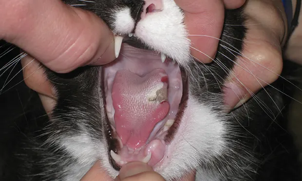
Electrical burn of the lateral aspect of a cat’s tongue caused by the cat chewing on an electrical cord.
Noncardiogenic pulmonary edema (NCPE) occurs secondary to systemic inflammation or neurogenic stimulation. NCPE is thought to develop after a massive catecholamine release and subsequent elevation in pulmonary capillary pressure and microvascular permeability. Underlying disease processes include seizures, traumatic brain injury, electrocution, systemic inflammatory response syndrome (SIRS), sepsis, and upper airway obstruction.

Electrical burn of a dog’s hard palate caused by the dog chewing on an electrical cord.
Clinical Signs
NCPE patients typically exhibit signs of respiratory distress, including tachypnea, dyspnea, and potential pulmonary crackles. Variability of underlying causes (eg, electrocution causing ulceration of the tongue and oral cavity [Figures 1 and 2]) necessitates a complete physical examination. Patient anxiety and respiratory distress may prohibit complete examination on presentation; if so, a partial examination can be performed before placing the patient in an oxygen cage and completing the examination.
Diagnosis
Diagnosis of NCPE is often based on history, examination, and radiographic imaging. Classic radiographic findings include increased interstitial or alveolar opacity, notably in the caudodorsal lung fields (Figure 3). This contrasts with radiographic findings in patients with cardiogenic pulmonary edema, notably perihilar edema. However, other forms of respiratory disease should be investigated, including cardiogenic pulmonary edema, pneumonia, and hemorrhage from trauma or coagulation abnormalities. Echocardiography can exclude cardiogenic edema and left-sided heart failure. A coagulation profile should be performed and pneumonia ruled out based on history, physical examination findings (eg, nonfebrile), and CBC results.


Lateral (A) and VD (B) thoracic radiographic views of a dog with NCPE. Note the focal interstitial to alveolar pattern present in the 3 caudodorsal lung fields.
Treatment
Treatment is supportive and primarily includes administration of supplemental oxygen. Additional therapy depends on the underlying cause (eg, antiepileptic drugs to control seizures, antibiotics and pain medication for oral ulceration from electrocution). Sedating analgesics (eg, morphine) may also be considered if the patient’s anxiety and stress is exacerbating respiratory distress.
In patients with NCPE, the massive catecholamine release can damage the pulmonary endothelium, increase pulmonary microvascular permeability, and cause protein and fluid to leak into interstitial and alveolar spaces. As a result, IV fluids must be used judiciously to prevent worsening of the pulmonary edema. Although furosemide use has been reported, there is not currently sufficient evidence to support standard use in NCPE patients. While furosemide can be considered to decrease bronchospasms and act as a bronchodilator, it has several risks, notably dehydration, as these patients frequently cannot tolerate high fluid rates (because of microvascular permeability and risk for worsening pulmonary edema). Furosemide would not be successful in removing this type of edema and may even remove hydrostatic fluid, leaving protein-rich material in the airways.
While underlying diseases may warrant specific therapy (eg, antibiotics for oral ulceration), evidence supporting the use of corticosteroids, bronchodilators, or antibiotics in patients with NCPE is lacking.
Prognosis
Prognosis for these patients depends on the primary underlying cause. For NCPE without significant comorbidities, the prognosis is excellent; most patients require 24 to 48 hours of hospitalization before discharge with no long-term ill effects.