No Tooth, No Problem...Really?
Sophie, a 5-year-old spayed female Maltese, was presented for evaluation of a fractured left maxillary fourth premolar tooth (#208).
HistoryThe owner had noticed that, despite eating and drinking normally, Sophie was chewing food only on the right side of her mouth. There were no other signs of oral discomfort or periodontal disease.
Diagnosis was a fracture of the left maxillary fourth premolar tooth (cause and duration unknown); the referring veterinarian began administration of meloxicam (0.1 mg/kg PO Q 24 H with food) 3 days before presentation at our hospital.
Physical ExaminationSophie weighed 3.1 kg and had a body condition score of 4/9. Findings on general physical examination, including heart rate, respiratory rate, temperature, chest auscultation, mucous membrane color, hydration assessment, abdominal palpation, and capillary refill time, were unremarkable.
Laboratory ResultsComplete blood count, serum biochemistry profile, and buccal mucosal bleeding time were within normal limits. (The latter test was performed to evaluate clotting time because extraction of the fractured tooth with pulp exposure was anticipated.)
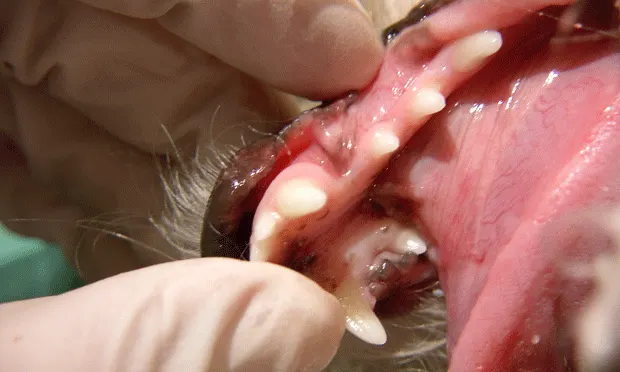
Oral ExaminationOnce the patient was under general anesthesia, oral examination revealed a complicated crown/root fracture of #208. The left and right mandibular first premolar teeth (#305 and #405, respectively) were noted to be clinically missing (Figure 1). All periodontal probing depths were normal except on the vestibular surface of #208, where the crown/root fracture was located.

Figure 1: On oral examination, teeth #305 (arrow) and #405 (arrowhead) were clinically missing.
Diagnostic Imaging
Full-mouth dental radiographs were taken (Figures 2 and 3).

Figure 2: Dental radiograph of the right rostral mandible

Figure 3: Dental radiograph of the left rostral mandible
ASK YOURSELF…
What is your tentative diagnosis, how would you confirm, and what treatment would you pursue?A. Missing #305 and #405: No further treatment or diagnostic testing neededB. Missing #305 and unerupted #405, with suspected dentigerous cyst: Surgical extraction of #405 and sampling of the cyst lining for histopathologic confirmation.C. Missing #305 and suspected abscess involving #405: Surgical extraction of #405 and cytologic examination of the exudate within the area of radiolucencyD. Missing #305 and suspected odontogenic tumor (ameloblastoma) at right mandible caudal to #404: Incisional or excisional biopsy; if ameloblastoma is confirmed, partial right rostral mandibulectomyE. Unerupted #305 and missing #405: No treatment necessary at this time; continue to monitor every 12 months with dental radiographs
CORRECT ANSWER:B. Missing #305 and unerupted #405, with suspected dentigerous cyst: Surgical extraction of #405 and sampling of the cyst lining for histopathologic confirmation
DiagnosisTentative diagnosis was an unerupted #405, with a dentigerous cyst forming around the crown, and a missing #305. Confirmation of this diagnosis is based on histopathologic examination of the cyst lining.
TreatmentAfter right caudal mandibular nerve block and gingival flap elevation, the soft tissue, fluid-filled cyst and unerupted #405 were exposed. The cyst was incised, fluid evacuated, and lining carefully removed by curettage. Tooth #405 was gently elevated, alveolus curetted, radiograph taken, and gingival flap closed with absorbable sutures. A postoperative dental radiograph was also obtained (Figure 4).
Additional DiagnosticsThe entire cyst lining was submitted for histopathologic examination. Histopathologic evaluation confirmed an epithelial lining consistent with a dentigerous cyst.
OutcomeAt 1-month follow-up examination, the gingival mucosa had healed over the surgical site and the sutures had dissolved. The surgical site of the cyst was expected to fill in with new bone; radiographs were recommended at the next dental cleaning. Sophie recovered well and returned to her normal activities.
DiscussionTooth Crown DevelopmentDuring embryologic development of the tooth crown, the outer and inner enamel epithelium condense to ultimately contribute to the formation of the enamel. Residual components of this structure cover the crown until eruption.1 As the tooth erupts, the crown penetrates these tissues through the gingiva, and normally these tissues form the epithelial attachment just apical to the cementoenamel junction.

Figure 4: Postoperative radiograph after surgery to remove dentigerous cyst and tooth #405
If the tooth does not erupt, the epithelial tissue creates a sac that retains the ability to produce fluid and the potential to form a true epithelium-lined cyst. Not every unerupted tooth develops into a dentigerous cyst, but which unerupted teeth form a cyst and when this occurs are unknown.
Dentigerous CystsThe source of fluid accumulation is debatable; proposed mechanisms include an osmotic gradient difference from within the cyst2 or an inflammatory response.3 In either case, the pressure of the fluid causes the bone to remodel around the expanding cyst, allowing these cysts to take up large portions of the jaw. The jaw integrity can be weakened, making it prone to pathologic fracture (as well as cystic expansion), which can potentially damage surrounding neurovascular tissues.4
Early detection is very important. If a tooth is unerupted and there is no evidence of cyst formation, extraction of the unerupted tooth should resolve any potential problem; however, yearly survey radiographic monitoring is another option.4 Histopathologic examination of the entire cyst lining is also important because some odontogenic tumors can clinically and radiographically mimic a dentigerous cyst but require more aggressive treatment.
Missing TeethMissing teeth are more often found in brachycephalic breeds and most commonly involve the first premolar teeth.5,6 Radiographs should be taken for every missing tooth. Finding an unerupted tooth with or without an associated cyst is an important finding that could otherwise go unnoticed, thereby allowing significant structural changes to the jaw bone before outward clinical signs are noted.
Case Synopsis
In this case, the initial presentation was for a fractured tooth (and associated discomfort), but careful oral examination, coupled with full-mouth dental radiographs, helped diagnose and quell another potential problem—the dentigerous cyst.
The original problem, the fractured tooth, was evaluated for endodontic therapy, but because of the extent of subgingival fracture and the thin remaining clinical crown, this tooth was extracted.