Nasal Discharge in a Dog
A 12-year-old spayed female English springer spaniel was presented for nasal discharge and intermittent epistaxis.
History
The dog is an indoor pet that only goes outside while on a leash. Two years ago, the dog had trauma to the left eye; she underwent enucleation and had a prosthetic globe placed.
Two months ago, a mucoid nasal discharge from the right nostril was noted. A 10-day course of antibiotics (amoxicillin/clavulanate, 13.75 mg/kg PO Q 12 H) was prescribed by the local veterinarian and the discharge resolved.
Two weeks ago, the owner noticed that the discharge had recurred and was now hemorrhagic. In the past week, the owners noted 2 episodes of mild epistaxis that resolved without treatment. The owners reported no coughing or sneezing and a normal appetite and activity level.
Physical Examination
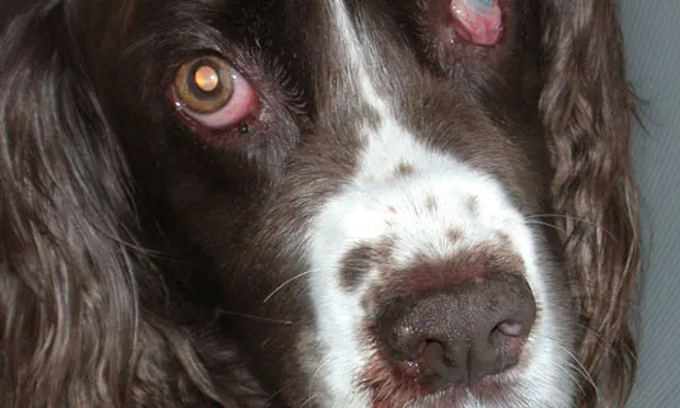
The dog was bright, alert, and hydrated, with a body weight of 19.6 kg. A hemorrhagic, mucoid discharge was noted coming from her right nostril (Figure 1). Airflow was absent from the right nostril. Her right eye retropulsed normally. Oral examination was unremarkable. Her submandibular lymph nodes were 0.5 cm in diameter. The rest of her physical examination, including blood pressure and temperature, was unremarkable.
Diagnostics
A CBC, serum biochemical profile, and urinalysis showed no abnormalities. Prothrombin time and partial thromboplastin time were normal. Thoracic radiographs were unremarkable. Lymph node aspirates from both submandibular lymph nodes revealed mild reactivity. Computed tomography (Figure 2) was done, followed by rhinoscopy using a rigid scope for obtaining biopsies.

ASK YOURSELF...
Besides a nasal tumor, what are other differentials for a mucoid or hemorrhagic nasal discharge?
What staging tests should be done for dogs with nasal tumors?
What are the main tumor types found in dogs with nasal tumors?
DID YOU ANSWER...
The most common local causes of nasal discharge in dogs besides cancer are allergic rhinitis, foreign bodies (such as plant material), fungal infections, oral nasal fistulas, parasites, and tooth root abscesses or other dental disease. Systemic diseases that may also lead to nasal discharge include coagulopathies, hypertension, hyperviscosity syndrome, and pneumonia.
The staging system for nasal tumors in dogs takes into account the extent of the local tumor, including bone and surrounding tumor invasion as well as nodal and distant metastasis. Minimally, staging tests should include thoracic radiographs, lymph node aspiration, and CT. Other tests to evaluate the general health of the patient and help rule out other diseases include CBC, serum biochemical profile, clotting profile, urinalysis, and abdominal ultrasound.
Carcinoma is the most common nasal tumor histology in the dog and includes squamous cell carcinoma, transitional cell carcinoma, adenocarcinoma, and undifferentiated carcinoma; of these, adenocarcinoma is most common, whereas squamous cell and transitional cell carcinomas are more rare. Sarcomas are the next most commonly seen type of nasal tumor and include chondrosarcoma, osteosarcoma, and fibrosarcoma. Less common types of tumors found include hemangiosarcoma, mast cell tumor, melanoma, plasma cell tumor, and lymphoma.
Diagnosis: Nasal Adenocarcinoma
CT (Figure 3) revealed a large, expansile mass within the right nasal cavity, with destruction of the frontal and lacrimal bones (green arrow) and extension into the right orbit (yellow arrow) and frontal sinus (red arrowhead). There was also extension of the mass into the right nasopharynx (green arrowhead). Extensive turbinate destruction was identified.

Computed Tomography
CT is usually the preferred imaging modality for suspected nasal tumors. Scans obtained both with and without contrast enhancement should be done because different information can be obtained from each study. The noncontrast study should be done first to evaluate the bone and nasal turbinates for destruction. Thin (1 mm) slices should be done through the area of the cribriform plate tolook for bone lysis and invasion into the olfactory bulb of the brain.
The contrast scan can help differentiate soft tissue (tumor) from mucus since tumors are better visualized with enhancement. Further, since the meninges will often contrast enhance, this strategy can be helpful to determine if there is tumor invasion into the brain, which can carry a worse prognosis. All imaging should be completed before rhinoscopy is done or blind biopmasies are taken because bleeding during the procedure can complicate image interpretation.
Rhinoscopy & Biopsies
A thorough oral examination should be performed. If possible, visualize the oropharynx by retroflexing a flexible endoscope to look for masses. The examination should also be done before biopsies are taken because there will be bleeding after the biopsy procedure that may obscure visualization of the area.
Biopsies can be taken from the nasal cavity with either a flexible or rigid scope. Blind biopsies can be obtained, although they are less likely to result in a diagnostic sample because tumor areas are not visualized before sampling. To prevent damage to the cribriform plate, take care not to obtain biopsies deeper than the medial canthus.
CT can be used to guide what areas to biopsy by showing the location of the mass lesion in the nasal cavity. This can be particularly helpful if it is difficult to visualize the mass, as can happen when there is epistaxis.
Treatment & Outcome
Radiation therapy is the most effective treatment for dogs with nasal tumors, and it is now available at many private and academic referral hospitals. The extent of the tumor as diagnosed on CT scan will determine what normal tissues are included in the radiation field. This will, in turn, determine side effects seen. The job of the radiation oncologist is to ensure maximal coverage of the tumor with the prescribed dose of radiation while minimizing dose to normal surrounding tissue. For example, if the eyes are in the radiation field there is a high chance that the dog will develop a cataract within one year. To help avoid this, advanced imaging and computerized treatment planning are necessary.
Prognosis
Median survival time with treatment ranges from 8 to 15 months for dogs with carcinomas. Chronic nasal discharge and occasional epistaxis may be present even after successful therapy.