Managing Maggots & Bots in Dogs & Cats
Jennifer E. Thomas, DVM, DACVD, Oklahoma State University
Mason V. Reichard, MS, PhD, Oklahoma State University Center for Veterinary Health Sciences
Myiasis, the infestation of live vertebrate host tissues with dipterous (ie, fly) larvae,1 is commonly called “maggot” or “bot” infestation and requires prompt veterinary treatment. Bot flies can invade skin; matted, soiled hair coats; or deeper tissues of dogs and cats. The fly larvae feed on the host’s dead or living tissues, body fluids, or ingested food, and lesions with a characteristic putrid odor can occur. Infestation can occur in the eyes, ears, nasal and oral cavities, skin, alimentary tract, urethra, and genitals.2
Myiasis is characterized as obligatory, facultative, or accidental, based on species‒host dependence2:
Obligatory myiasis is caused by flies whose larvae require a living host for development. Cochliomyia hominivorax (ie, primary screwworm) and all bot flies are classic examples of flies that cause obligatory myiasis. In cats and dogs, the most commonly encountered bot flies are Cuterebra spp (eg, rodent bot fly, rabbit bot, warble).3 Flies causing obligatory myiasis may lay eggs in uninfected wounds that may be as small as a tick bite (C hominivorax) or penetrate through the skin of their hosts to invade healthy tissues (Cuterebra spp). Yorkshire terriers appear to be at higher risk of Cuterebra spp infestation than other breeds.4
Facultative myiasis results when nonparasitic flies opportunistically infest dogs and cats. These flies usually lay eggs in decomposing organic matter (eg, carrion, feces) but may deposit eggs in animals with infected, open wounds, or hair coats soiled with feces, urine, or vomitus. Larvae typically feed on dead or decaying host tissue. Facultative myiasis in soiled or contaminated wounds most often is caused by species of Calliphora (ie, blow flies), Lucilia (ie, green bottle flies, blow flies), Musca (eg, M domestica, the common house fly), Phaenicia (ie, green bottle flies, blow flies), Phormia (ie, black blow flies), and Sarcophaga (ie, flesh flies).2,5 Dogs and cats with access to the outdoors and animals with weakened immune systems have a greater risk for infestation. Obesity in cats has been identified as a predisposing factor for infestation with Calliphora erythrocephala.6
Accidental myiasis occurs when dogs and cats ingest fly eggs or larvae. Accidental myiasis is pseudoparasitism because further life-stage development of these larvae does not occur in the GI tract. Immature stages typically are eliminated without causing clinical disease.2 Fly eggs and larvae are found in vomitus or feces.
Diagnosing Myiasis
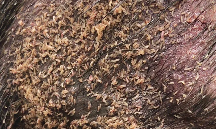
Affected dogs and cats typically are presented because of macerated or erosive/ulcerated skin lesions in which fly larvae are often visible. The lesions may be covered by thickly matted hair and are often soaked in wound exudate, so clients are likely unaware of the extent of the cutaneous lesions or the presence of fly larvae. The infested lesions often emanate a putrid odor.7,8 In cases of Cuterebra spp infestation, dogs and cats present with large swellings of the skin and subcutaneous tissue with a large central opening through which larvae may protrude.
Recovery and identification of fly larvae from infested animals is key to diagnosis. Larvae may be recovered from lesions, feces, or vomit, depending on myiasis type. Identification is based on the morphologic structure of larval mouthparts, spiracles (ie, the opening of the respiratory system), and arrangement of cuticular spines.9 Larvae of myiasis-causing flies are either maggots or bots.2
Maggots are long and slender and typical of Calliphora, Cochliomyia, Lucilia, Musca, Phaenicia, Phormia, and Sarcophaga spp. Bots are large and rotund (eg, Cuterebra spp). (See Figure 1.) Third-stage larvae of C hominivorax contain pigmented tracheal trunks (see Figure 2) that aid in differentiation from other myiasis maggots. Third-stage Cuterebra spp larvae are found most often in subcutaneous cysts in dogs and cats. (See Figure 3.) Cuterebra larvae are notable by their large size (ie, ≈2.5 cm long) and dark color, resulting from the stout, black spines covering the bot body.

FIGURE 1
Overall morphology and spiracular plates of third-stage larvae of common obligatory and facultative myiasis-causing flies in dogs and cats. A. Cuterebra spp, B. Cochliomyia hominivorax, C. Phaenicia spp, D. Phormia spp, and E. Musca spp. Size bar = 30 mm.
Figure 1 courtesy of Mason Reichard, MS, PhD, and Megan Wholtjen, National Center for Veterinary Parasitology, Oklahoma State University
Dogs and cats are accidental hosts for Cuterebra spp larvae, which can be found migrating through a variety of tissues,10-19 including the eyes and central nervous system, with deleterious effects. Many diagnostic laboratories also perform larval identification, which can be valuable in regions of increased risk for reportable forms of myiasis. C hominivorax infestation in any animal species should be reported to the US Department of Agriculture (USDA).20
Laboratories Offering Larval Identification
The following are some of the diagnostic laboratories that offer larval identification at the time of publication:
Antech
Cornell University
IDEXX
National Veterinary Services Laboratories
Oklahoma State University
Texas A&M University
University of Florida
University of Georgia
University of Prince Edward Island, Canada
University of Tennessee
Treating Infestation & Myiasis
Infestation of myiasis-causing fly larvae in dogs and cats requires immediate veterinary attention and therapy. Assessment and treatment frequently require sedation or anesthesia of the infested patient.5,21
Infested wounds should be clipped to facilitate full assessment and cleaning of lesions. Mechanical removal of larvae with surgical excision and aggressive debridement of necrotic tissues will likely be required.22 Chlorhexidine and povidone-iodine are recommended for wound cleaning and lavage.23
Rupture of Cuterebra spp can be accompanied by severe reaction, including anaphylaxis, and care should be taken to remove the larvae whole. The breathing pore for Cuterebra spp is often much smaller than the spine-covered larvae, and the opening should be surgically enlarged to allow for intact extraction. (See Figure 3.)
Antiparasitic compounds are often administered before, or shortly after, mechanical removal of larvae to kill remaining organisms. Historically, pyrethrin-containing compounds were applied to infected sites of dogs only.7,24 Current antiparasitic compounds with reported efficacy for treating and controlling myiasis in dogs and cats include ivermectin,6,23,25 nitenpyram,7 and spinosad with milbemycin.26 Currently approved isoxazolines (eg, fluralaner, sarolaner, afoxolaner) also appear to be safe and effective at the doses on the labels.24
Secondary infections of these lesions are common, and the patient should be placed on appropriate antibacterial or antifungal compounds for a minimum of 14 days beyond clinical resolution. The presence of bacterial or fungal microorganisms should be demonstrated by cytology prior to initiating systemic therapy. Post-treatment, clients should be instructed on proper wound management.
Preventing Myiasis
Preventing myiasis in dogs and cats includes keeping wounds clean, treating underlying medical conditions, housing weak and debilitated patients indoors or in screened enclosures, and maintaining a healthy hair coat free of mats, urine, and feces.
Conclusion
C hominivorax was eradicated in the United States in 1966, reintroduced during isolated outbreaks, and re-eradicated in 2016. (See History of Primary Screwworm.) However, the re-emergence of C hominivorax in the U.S., and the constant threat of introduction by dogs and cats that travel abroad, highlight the need for veterinarians to remain continually vigilant for rare and foreign animal diseases. Recognizing the signs of larval infestation and initiating treatment promptly are essential to ensure animal health and prevent the spread of disease.
History of Primary Screwworm
C hominivorax in the United States has been eradicated, reintroduced, and re-eradicated.
C hominivorax was successfully eradicated from the United States in 1966 and from Mexico in 1991.27 Eradication was achieved through a program operated by the USDA employing the release of sterile male flies.
More recently, C hominivorax was reintroduced into the U.S. in isolated incidents among animals and humans after travel to endemic areas.28
On October 3, 2016, the USDA’s Animal and Plant Health Inspections Service announced infestations of C hominivorax among Key deer on Big Pine Key, Florida, in the National Key Deer Refuge.29
Infestation with C hominivorax was reported in approximately 135 Key deer, 3 dogs, 2 cats, 1 pig, and 1 raccoon.30
Containment and re-eradication was achieved through the release of 154 million sterile flies, 17 000 animal inspections, and approximately 700 hours of surveillance.30
This outbreak was contained, and primary screwworm has again been eradicated from the U.S.
This article originally appeared in the May 2018 issue of Veterinary Team Brief.