Managing Chronic Maladaptive Pain
HISTORYAccording to the previous owner, Payton had started limping on the right rear limb about 1½ years before surrender. No diagnosis or treatment had been pursued. Payton’s diet at presentation consisted of a commercial dry dog food.
PHYSICAL EXAMINATIONPayton weighed 47 kg, had a body condition score of 3/5, and exhibited grade 4/5 right rear limb lameness with dramatic atrophy of the right thigh muscles. The left thigh circumference was 46 cm, while the atrophied right thigh measured 40 cm.
Related Article: Handling Pathologic Pain
Systematic pain palpation (see Pain Examination by Palpation)1 revealed moderate pain at the thoracolumbar junction, iliopsoas muscle bundle, and proximal quadriceps, and Payton objected to full extension or hyperextension of the right stifle. Based on the visual analog scale (VAS), the overall pain score was 5/10. (While the VAS is not objectively validated if multiple observers assess pain, it can be a consistent vehicle for tracking pain if initial and follow-up assessments are done by the same clinician.)
Significant buttressing of the proximal right tibia with palpable thickening of the stifle joint capsule was noted, along with mild medial buttressing of the proximal left tibia. No apparent effusion was evident. Under sedation, the right stifle demonstrated positive anterior drawer and tibial thrust. The left stifle demonstrated subtle anterior drawer in partial flexion but not in extension. These findings were consistent with bilateral cranial cruciate ligament (CCL) disease, and Payton was diagnosed with complete CCL rupture of the right knee and partial tear of the left.
Related Article: Pain Management in CatsLABORATORY FINDINGSComplete blood count, serum biochemical profile, and thyroxine test results were normal. An E.R.D.-HealthScreen (heska.com) was negative for microalbuminuria (urine specific gravity, 1.030).
Ask Yourself...what initial plan is most appropriate for Payton?
A. Schedule surgery immediately to explore the right stifle and perform tibial plateau leveling osteotomy
B. Prescribe short-term pain relief and schedule stifle surgery in 2 weeks.
C. Devise a strategy to control pain, support and improve joint health, and restore function and strength.
Correct answer: C. Devise a strategy to control pain, support and improve joint health, and restore function and strength.
WHAT IS MALADAPTIVE PAIN?Clifford Woolf of Harvard Medical School is credited with articulating the concept of adaptive versus maladaptive pain. According to Woolf, “adaptive pain contributes to survival by protecting the organism from injury or promoting healing when an injury has occurred.”2 He identifies maladaptive pain, by contrast, as “an expression of the pathologic operation of the nervous system; it is pain as disease”2 (ie, pain that does not serve a useful physiologic purpose and is the result of nervous system changes that facilitate the pain experience). Woolf also identifies the need to provide targeted therapy, focusing on specific pathologies contributing to pain as well as specific receptors in the nervous system to achieve optimum outcomes.2
When a patient with CCL disease and stifle osteoarthritis has persistent lameness that leads to generalized maladaptive caudal back pain, management of multiple issues is required to achieve optimum clinical results. Physical medicine and rehabilitation techniques provide a route to restore function and strength; however, it is ethically questionable to manipulate painful tissues in a nonverbal individual without first effectively reducing pain. In addition, physical medicine techniques alone, including surgical stabilization, do not address the nervous system remodeling (particularly in the dorsal horn) that occurs with chronic pain.2
Related Article: Prevent Chronic Pain By Treating Acute Pain First
PAIN PROTOCOLSTo target the local, chronic inflammatory component of Payton’s pain, the dog was treated with meloxicam (1.5 mg/mL suspension) at 0.1 mg/kg PO Q 24 H as well as gabapentin at 8.5 mg/kg PO Q 12 H, thereby targeting the maladaptive pain component by altering calcium permeability in the dorsal horn neurons by way of the alpha-2-d ligand, raising the threshold for neuronal depolarization.3 Polysulfated glycosaminoglycan (PSGAG, 100 mg/mL SC, Adequan Canine, novartis.com) is not approved by the U.S. Food and Drug Administration for treatment of CCL disease but was administered because of its disease-modifying effects in osteoarthritis, an inevitable sequela in CCL disease.3 PSGAG was given at 4.4 mg/kg SC twice weekly for 4 weeks, once weekly for 4 weeks, and twice monthly for ongoing maintenance.NUTRITIONAL THERAPYObesity contributes to the prevalence and progression of osteoarthritis; therefore, Payton’s body condition score of 3/5 was optimal to allow the nutritional therapy to focus exclusively on joint support.3 Payton was fed a veterinary therapeutic ration formulated specifically for joint support; this diet has been demonstrated to improve function in dogs with osteoarthritis.4
Pain Examination by Palpation
Palpate the affected area with approximately 4 kg of pressure using the fleshy surface of the third phalanx of the index and second fingers—not the tips of the fingers (use enough pressure to deform the tissue and blanch out the end of the fingernail). This is the digital palpation technique used by human pain practitioners to identify tender points to assist in the diagnosis of fibromyalgia.1
Perform palpation systematically and the same way each time:
Palpate the paraspinal musculature at approximately each spinal segment from occiput to sacrum.
Palpate the circumference at the base of the neck by the hands of the clock—10 & 2, 9 & 3, 8 & 4.
Palpate caudal to the scapula by the hands of the clock—10 & 2, 9 & 3, 8 & 4.
Palpate at the thoracolumbar junction by the hands of the clock—10 & 2, 9 & 3, 8 & 4.
Palpate the lateral lumbar muscles by spinal segment.
Palpate the iliopsoas muscle bundle starting at the thoracolumbar junction and proceeding to the pubis.
Squeeze the proximal quadriceps.
Perform joint range-of-motion from toes to torso.
Return to areas generating a reaction and evaluate with additional palpation and/or range-of motion manipulations to better identify and characterize the presence and nature of pain.
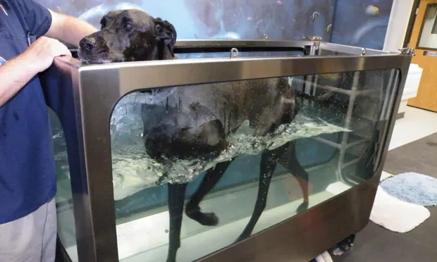
PHYSICAL REHABILITATIONPayton was evaluated for pain and function every 2 weeks for 1 month, at which time her overall VAS score had stabilized at 1/10 and the right rear limb lameness was reduced to grade 1/5. Physical rehabilitation was then initiated to improve function and build strength. Therapy sessions using the underwater treadmill were designed to help build strength while minimizing discomfort. Payton was buoyed by the water, allowing exercise in a biomechanically normal posture while minimizing weight bearing on the joints. In addition, it is harder to move through water than air, so the increased resistance strengthens muscles.3 Sessions were timed at 20 minutes twice weekly for 8 weeks. The water measured 18 inches (reaching Payton’s mid-thigh as shown in Figure 1) and treadmill speed ranged from 1.8 to 2.25 mph.

Figure 1. Payton underwent twice-weekly 20-minute hydrotherapy sessions at treadmill speeds of 1.8 to 2.25 mph. Note that the water measures about 18 inches.
The owners had restricted Payton’s walks to 10 to 15 minutes once or twice a day. As the pain score decreased and her strength increased, the length of at-home walks was increased by 10 minutes per walk every 2 weeks to a maximum walk time of 60 minutes. By week 16, Payton’s overall VAS score on palpation was 0/10.
LASER THERAPYLaser therapy (980 nm, 6W, CW, 20 J/cm2 delivered at the surface of the skin, Figure 2) was also instituted at the same time as physical rehabilitation to address persistent pain at the thoracolumbar junction. Therapeutic laser treatment provides photobiomodulation at a cellular level and has been used for the treatment of multiple musculoskeletal issues, including osteoarthritis.3
Figure 2a. Payton received 4 weekly laser treatments at the thoracolumbar junction and the iliopsoas muscles; Figure 2b. Close up
OUTCOMEEight weeks after initiation of multimodal pain management, the interval between Payton’s reassessments was increased to monthly. At the 3-month mark, the meloxicam dose was decreased to 0.05 mg/kg PO Q 24 H, then 1 month later to 0.025 mg/kg PO Q 24 H, then discontinued the following month. At this point, the gabapentin dose was decreased as well and pain reassessed every 8 weeks. Gabapentin was decreased slowly to avoid a pain rebound5: first to a morning dose of 400 mg PO and an evening dose of 200 mg PO for 8 weeks, then 200 mg PO Q 12 H for 8 weeks, then 200 mg PO Q 24 H for 8 weeks, then discontinued.
Payton’s ongoing maintenance includes a therapeutic joint support diet; PSGAG (4.4 mg/kg) SC twice monthly; and glucosamine, low-molecular-weight chondroitin, and avocado-soybean unsaponifiables (ASU, added at age 7 years). The dog displays full activity, walking 12 to 20 miles per week, receives no pain medication, and no longer exhibits any right rear limb lameness. The overall VAS score remains 0/10. Surgery of the right stifle was avoided, and the dog has no apparent additional degeneration of the left stifle.
CLOSING REMARKSThis case illustrates the utility of a multimodal approach to chronic maladaptive pain and the potential for return to full function. Other pharmaceuticals (eg, amantadine, tramadol) and physical medicine modalities (eg, acupuncture, medical massage, trigger point therapy) were available for use if needed.
At continuing education meetings, veterinarians and veterinary healthcare teams are now consistently exposed to strategic multimodal management of chronic pain. Because each patient presents a unique set of signs, determining which modalities are best can be challenging. For patients with CCL disease, such as Payton, when surgery is not an option, a return to full comfort and function is still possible.

Treating patients with chronic, maladaptive pain can be extremely rewarding because these cases challenge the veterinary healthcare team to continue expanding treatment options as new information becomes available and to restore the famiily-pet relationship when it has been undermined because of pain and dysfunction.
The final patient plan may include surgical intervention, depending on the patient’s response to therapy and the owner’s wishes and financial resources.
TAKE-HOME MESSAGES
Every patient in pain has unique needs and nuances.
Chronic pain can be maladaptive and warrants a multimodal approach.
Patients with chronic, maladaptive pain need and deserve regular reassessments so their “pain plans” can be adjusted.
In accordance with the American Animal Hospital Association/American
Association of Feline Practitioners Pain Management Guidelines for Dogs and Cats, thoroughly assess and treat all diagnosed issues at each visit.
Once chronic maladaptive pain is reduced or controlled, strive to improve the patient’s function and strength.
Control pain, improve function, build strength, and then decrease pain medication to the lowest effective dose (which may be zero).
CCL = cranial cruciate ligament, VAS = visual analog scale, ASU = acocado-soybean unsaponifiables, PSGAG = polysulfated glycosaminoglycan
MANAGING CHRONIC MALADAPTIVE PAIN • Robin Downing
References
1. The manual tender point survey. Sinclair D, Starz TW, Turk DC. http://www.fmaware.org/News2eb58.html?page=NewsArticle&id=6263; reprinted from FMOnline Oct 2005; accessed July 2011.
Pain: Moving from symptom control toward mechanism-specific pharmacologic management. Woolf CJ. Ann Intern Med 140:441-451, 2004.
Multimodal Management of Canine Osteoarthritis. Fox SM, Millis D—London, UK: Manson Publishing, 2010.
A multicenter study of the effect of dietary supplementation with fish oil omega-3 fatty acids on carprofen dosage in dogs with osteoarthritis. Fritsch DA, Allen TA, Dodd CE, et al. JAVMA 236:535-539, 2010.5. Information by drug name: G drugs. Veterinary Anesthesia and Analgesia Support Group. http://www.vasg.org/g_drugs.htm; updated June 2011, accessed July 2011.