Malocclusion in the Rabbit
You have asked…What do I need to know to best diagnose and treat malocclusions in a rabbit?
The expert says…
Rabbits (herbivores) masticate a large volume of fibrous food material in their lifetime. They have continuously growing incisors and cheek teeth with relatively long crowns, only parts of which are visible within the oral cavity. Subgingival reserve crown erupts into the oral cavity as dental wear dictates. With advanced age, germinal tissue finally forms the definitive root and ceases making a crown; at this time, remaining subgingival reserve crown erupts and is worn away, leaving a gap in the dental arcade.
If conditions alter the balance between tooth wear and formation, dental disease may result, and if left unrecognized and/or untreated, can become life-threatening.
Related Article: Brief Overview of Dental Disease in Pet Rabbits
Diagnosis
Skeletal & Dental Malocclusions

Domestication has resulted in the creation of new breeds of rabbit, with dolichocephalic (Figure 1), brachycephalic (Figure 2), and mesaticephalic (Figure 3) head types.1 Brachycephalism, with its associated domed forehead and shortened maxilla, and especially when combined with dwarfism, predisposes to class III (skeletal) malocclusions.
Figure 1. Dolichocephalic head type

A class III malocclusion leaves the first mandibular cheek tooth and the last maxillary cheek tooth without an opposing tooth against which to chew, possibly resulting in inappropriate wear. Overgrowth of these crowns effectively lock the cheek teeth arcades and prevent all rostral or caudal chewing motions that may have previously existed, further preventing crown wear.
Figure 2. Brachycephalic head type with dwarfism
Other dental malocclusions can occur from a displaced tooth bud or from insufficient space or abnormal forces during eruption, which displace a tooth out of the arcade. These unopposed individual teeth overgrow, causing soft-tissue trauma.
Acquired Malocclusion
An acquired malocclusion in herbivores is usually secondary to insufficient wear of teeth from poor feeding practices. Ongoing crown production and eruption without equivalent wear results in excess crown length within the oral cavity. As the maxillary and mandibular cheek teeth erupt, the jaws are forced open until they cannot open any further. Teeth then start to tip over into abnormal orientations. Tipping deforms the crestal alveolar bone on the side that the tooth is tipping toward, causing gaps between the tooth and alveolus on the side from which the tooth is moving away.
If conditions alter the balance between tooth wear and formation, dental disease may result, and if left unrecognized and/or untreated, can become life-threatening.
These gaps become filled with food and debris that act as wedges to further accelerate tipping and also allow apical movement of bacteria, which can cause infection. When excess crown length can no longer be accommodated within the oral cavity, germinal tissue and apices of the teeth are forced retrograde into the alveolar bone. In severe cases, the teeth breach the ventral cortex of the mandible as well as invade the maxillary sinuses and retrobulbar spaces. This overgrowth is painful as apical pressure crushes the nerves and deforms the alveolar bone.
Depending on the personality of the patient and the astuteness of the client, behavioral changes from this pain may be detected early enough in the process to direct treatment before oral disease becomes more severe. Often, however, patients are not presented until a palpable bony deformation is detected along the mandible or abscessation has occurred.
Related Article: Anorexia & Lethargy in a Rabbit
Treatment
Primary Incisor Malocclusions
Primary incisor malocclusions are uncommon. Almost all incisor malocclusions are secondary to abnormalities in the cheek tooth occlusion.2 Any animal presented with an abnormal incisor occlusion must be assessed for cheek tooth disease before a primary incisor malocclusion can be assumed. Treatment should be directed at correcting the cause. If the cause cannot be corrected, care involves either lifelong intermittent crown reduction or extraction.

Cheek Teeth Malocclusions
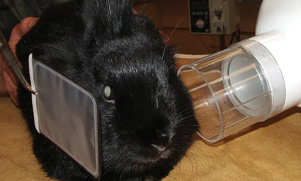
Determining the presence and extent of cheek tooth disease is the first step in treatment. An intraoral examination via endoscopy or otoscopy only detects intraoral overgrowth. Skull radiographs are essential to accurately determine the extent of oral disease. Radiographs of a smaller, compliant patient can be taken with a dental x-ray machine without sedation (Figure 3), but large and/or agitated patients require sedation for acquisition of skull films using a standard x-ray table. Typically, right and left lateral, right and left lateral-oblique, and dorsoventral views are required. Once acquired, interpretation can be facilitated using anatomic reference lines.3
Figure 3. In a small, compliant patient, dental radiographs may be taken without sedation: Lateral radiograph (A), dorsoventral radiograph (B)
Full anesthesia is necessary to access the oral cavity. Multiple protocols to safely anesthetize a rabbit exist, but intubation is preferred for prolonged procedures. Treatment begins with reduction of the intraoral crown length to more anatomically correct contour(s). Probing helps detect periodontal pockets; these should be cleaned. Any tooth with deep pockets, mobility, and purulent debris is a candidate for extraction, especially if an abscess is present. Once the diseased tooth is extracted, the abscess should be opened from the dermal aspect, cleaned, debrided, and flushed copiously. Suturing the stoma open slows closure, allowing treatment until the infection is resolved.
Related Article: Surgical Treatment of Periapical Mandibular Abscess in Rabbits
Antibiotics & Diet
Most antibiotics safe for oral dosing in rabbits do not cover an adequate spectrum to eliminate bacteria typically cultured from an abscess, making it necessary to teach owners how to give antibiotic injections at home. The abscess site should be reassessed 14 days post surgery, under sedation if needed, to ensure the site remains free of purulent debris. Follow-up sedated or anesthetized oral examinations are then scheduled to ensure intraoral crowns are maintaining a healthy length.
Address diet with clients to increase dietary fiber (ie, pellets and soft fruits are significantly limited, hay comprises the majority of the rabbit’s diet). Adequate calcium intake must also be ensured if a deficiency is suspected. Chew toys can be introduced to encourage oral exercise, and these can also provide enrichment benefits. Periodic professional oral examinations, especially for dwarf breeds, are recommended.
JUDY ROCHETTE, DVM, FAVD, DAVDC, provides referral dental and oral surgery services at West Coast Veterinary Dental Services in Vancouver, British Columbia, which she co-owns. Her articles have been published in Journal of Veterinary Dentistry, Clinician’s Brief, and Veterinary Clinics of North America. Dr. Rochette graduated from University of Guelph before becoming an Academy of Veterinary Dentistry fellow. A consultant for Veterinary Information Network, Dr. Rochette lectures locally and internationally, including at the NAVC Institute.