Leptospirosis in Dogs
George E. Moore, DVM, PhD, DACVIM (SAIM), DACVPM (Epi), Purdue University
Profile
Definition
Although leptospirosis was reported sporadically in the 1980s, it is now recognized as an important cause of acute renal disease in dogs as well as one of the most significant waterborne zoonotic diseases in the world.
Leptospira species are gram-negative bacteria.
Leptospira species are maintained in the kidneys of mammalian reservoir hosts. Leptospires are passed via urine from reservoir hosts and can contaminate ground water, which serves as the source of infection for other animals.
More than 200 serovars have been identified. Serovars Icterohaemorrhagiae and Canicola were prevalent in dogs in the 1950s to 1970s, but Grippotyphosa and Pomona have been dominant during the past 20 years.
Signalment
Originally, leptospirosis affected mostly sporting, working, or herding dogs with extensive exposure to the outdoors. Today, smaller breeds (including toy breeds) are diagnosed more frequently.
All ages and breeds are susceptible.
Geographic Distribution
Leptospirosis has been reported in all 50 states, with greater numbers noted in the Midwest, Northeast, and along the West Coast.
Climate may be responsible for major differences in prevalence internationally but probably causes only small variations in the U.S.
Prevalence estimates are greatly influenced by clinical suspicion of the disease and diagnostic testing choices.
At Risk
Unvaccinated dogs that come into contact with contaminated water sources are considered the most at-risk. Leptospirosis is also seen in rural and suburban areas with large areas of mammalian wildlife host habitats.
New infections are commonly seen in the autumn months because of increased wildlife movement and territorial marking. After heavy rains, areas of standing water increase, which can lead to greater exposure to all waterborne bacteria.
Pathophysiology
The helical shape of bacteria facilitates penetration of mucous membranes or broken epithelia (Figure 1).

FIGURE 1 The helical shape of leptospires facilitates the penetration of mucous membranes or broken skin.
Leptospires have an affinity for renal proximal tubules, the biliary tract, and vascular endothelium. They may also spread to the spleen, central nervous system, eyes, and genital tract.
Cellular damage from disease is most likely caused by leptospiral ipopolysaccharides and other components of the outer membrane.
Signs
Species differences in the host’s innate response to infection may impact disease severity.Mild disease consists of nonspecific illness, transient lethargy, and anorexia. Different patients manifest varying degrees of severity. Severe disease is typically associated with azotemia (eg, vomiting, dehydration, diarrhea, polyuria, and polydipsia; +/- oliguria and anuria).
Acute renal disease is most common, but acute-on-chronic renal failure is also seen.
Chronic renal failure has been documented as a sequel in 15% to 25% of dogs with leptospirosis that present with acute renal disease. Interstitial nephritis is common (Figure 2).

FIGURE 2 Interstitial nephritis is seen in many dogs with leptospirosis.
Jaundice secondary to cholangiohepatitis as well as anemia and hypoproteinemia secondary to vasculitis are present with severe disease.
Uveitis or bleeding tendencies, including dyspnea from pulmonary hemorrhage, are less common in dogs.
Although greater pathogenicity is suspected with some serovars (eg, Icterohaemorrhagiae), there are no studies following natural exposure to confirm this.
Feline Disease
Serologic evidence of feline disease has been noted, but clinical disease is rarely reported.
Experimental infection in cats generally results in mild disease despite the presence of renal and hepatic inflammation.
Rare reports of disease may be related to an innate immune response to recognize and phagocytize leptospires.
It is unknown whether cats can contaminate the environment with leptospires.
Diagnosis
Definitive Diagnosis
Definitive diagnosis is only possible after identification of the organism on histologic examination or through culture. Culture for Leptospira species is not ideal because it requires special media and has time constraints.
A 4-fold rise in microscopic agglutination test (MAT) antibody titers over 2 to 4 weeks is considered definitive. A single titer of 1:800 or greater in an unvaccinated dog with clinical signs suggestive of leptospirosis strongly supports diagnosis.
Lower titers may be seen in asymptomatic vaccinated dogs or asymptomatic dogs with a history of exposure.
Diagnostic Differentials
Diagnostic differentials include various causes of acute or chronic renal disease, cholangiohepatitis, vasculitis, uveitis, anemia, etc, due to toxins, septicemia, or other infectious diseases.
Lack of fever or leukocytosis should not eliminate leptospirosis from the list of differentials.
Laboratory Findings
Common findings include typical changes associated with renal disease. Azotemia (increased serum urea nitrogen and creatinine concentrations) and hyperphosphatemia are present with severe disease. Electrolyte changes may accompany azotemia and vomiting.
Hepatobiliary damage is indicated by increases in alkaline phosphatase and bilirubin concentrations. An increase in alanine aminotransferase concentration is possible, but not common.
Anemia, hypoalbuminemia, and leukocytosis are noted in some patients.
Imaging
No pathognomonic radiographic changes have been reported.
Renomegaly is often seen with acute renal failure.
Renal ultrasonography may show a hyperechoic pattern with loss of distinction of the corticomedullary border if vasculitis is present. Thoracic radiography may also show pulmonary changes caused by vasculitis.
PCR Assay
Polymerase chain reaction (PCR) testing may not be reliable for leptospirosis, as falsepositive and false-negative results are possible.
The test specific to Leptospira species is not conducted by all laboratories, and most PCR assays do not provide serovar or serogroup identification.
Necropsy
Gross lesions will vary with severity of infection and involvement of multiple organ systems. The kidneys are often enlarged and may have evidence of subcapsular or corticomedullary hemorrhage.
The liver may be enlarged and friable.
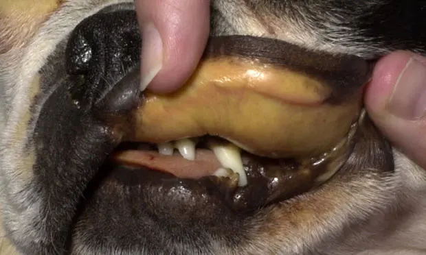
Icterus may be present (Figure 3), although congestion of mucous membranes with petechial or ecchymotic hemorrhage is sometimes seen.

FIGURE 3 Icterus due to biliary tract inflammation occurs in some cases of leptospirosis.
Treatment
Inpatient
Most patients require hospitalization for azotemia, renal disease, hepatic disease, etc.
Fluid terapy is indicated for dogs with organ damage and electrolyte or acid-base disorders.
Measurement of fluid intake (PO, IV) and urine output during fluid therapy is crucial because of variations in presentation (oliguria, anuria, polyuria, hemodynamic instability, etc).
Measurement of hepatic enzyme and serum protein concentrations, electrolytes, and acid-base disorders, as well as monitoring of azotemia, is recommended Q24 H during hospitalization.
Medications
In dogs, optimum treatment is extrapolated from human and laboratory animal studies instead of clinical trials.
Antibiotics are indicated to eliminate active infection and potential carrier states.
Doxyclycline or amoxicillin is the preferred oral medication.
Ampicillin (22mg/kg IV Q 6 H) or penicillin G potassium (25,000-40,000 Ueasu/kg IV Q 6 H) is recommended if oral medication is contraindicated. Dogs with oliguric or anuric renal failure should receive reduced doses of ampicillin because of decreased excretion of the drug.
Ampicillin or penicillin G must be followed by doxycycline (5 mg/kg PO Q 12 H) for at least 2 weeks to eliminate infection.
Doxycycline (5 mg/kg IV or PO Q 12 H) can be prescribed as initial treatment if vomiting or adverse reactions do not preclude administration.Optimal duration of doxycycline treatment is unknown, but administration should continue for at least 2 weeks after gastrointestinal signs resolve.
Ceftriaxone, cefotaxime, and azithromycin can be effective treatment for leptospirosis in humans, but their efficacy in dogs is unknown. Fluoroquinolones, chloramphenicol, and firstgeneration cephalosporins have questionable or no efficacy against leptospirosis.
Treatment at a Glance
To Eliminate Active Infection & Carrier State
Doxycycline or amoxicillin is preferred if oral medication is tolerated.
Ampicillin or penicillin G potassium IV can be administered if oral medication is contraindicated.
Follow with doxycycline for at least 2 weeks after gastrointestinal signs resolve.
Hospitalized Dogs With Azotemia or Renal/Hepatic Disease
Fluid therapy if organ damage and electrolyte or acid–base disorders are present.
Measurement of fluid intake (PO, IV) and urine output during fluid therapy is crucial.
In General
Prognosis
Leptospirosis can be fatal, even with appropriate antibiotic treatment.
Mortality rates of 10% to 30% are reported in dogs with moderate to severe organ damage, although many dogs exposed to leptospirosis have few or transient nonspecific clinical signs.
Prevention
Annual vaccinations for 4 serovars (Grippotyphosa, Pomona, Icterohaemorrhagiae, and Canicola) have high efficacy and are recommended. Vaccines for only 2 of the serovars generally do not provide cross-protection and are not recommended.
Zoonotic Risk
Patients should be considered a zoonotic risk to hospital personnel and other patients until at least 48 to 72 hours after initiation of antibiotic treatment.
At a minimum, hospital personnel should wear gloves when handling patients during this time. Face protection to prevent urine contact with eyes is also recommended. Urine should be considered infectious and disposed of accordingly until several days after initiation of antibiotic therapy.
Hospital cages should be marked appropriately.
Other dogs in the client’s household should be treated with doxycycline because of possible concurrent or common exposure.