Leak Testing After Jejunal Enterotomies
J. Brad Case, DVM, MS, DACVS, University of Florida
In the literature
Mullen KM, Regier PJ, Waln M, Colee J. Ex vivo comparison of leak testing of canine jejunal enterotomies: saline infusion versus air insufflation. Vet Surg. 2021;50(6):1257-1266.
The Research …
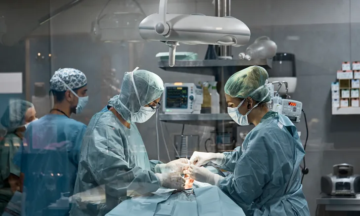
Accurate operative technique during intestinal surgery is key to avoid complications (eg, dehiscence, septic peritonitis) and should include handling atraumatic tissue, preserving the blood supply, placing sutures <2 to 3 mm between bites and >3 mm from the cut edge, snug primary apposition of wound edges, lack of mucosal eversion, and avoiding slack in the suture line.
Several intraoperative methods to predict the risk for intestinal dehiscence have been advocated for, with an intestinal leak test possibly being the most common. Although specific procedural steps have been reported, the main objective is to inject saline into the lumen of the surgical site while occlusive pressure is applied to the intestine around the surgical site. If saline is observed leaking through the enterotomy or anastomosis site, additional interrupted sutures or revision of the closure is recommended. Little evidence, however, exists in the veterinary literature to support or refute the validity of the leak test.
This ex vivo cadaveric study compared leak pressures between enterotomies tested with a saline leak test and an air leak test. Air leak tests are commonly performed in lower GI repair in humans but have not been reported in dogs. Results showed that canine intestinal leakage occurs at lower intraluminal pressures when tested with air than with saline, rendering leak testing more sensitive and precise with air as the infusate. This may suggest that detection of leakage at a lower pressure can improve sensitivity (ie, reduced chance of false-negative results) and reduce the risk for dehiscence; however, no clinical investigation has been performed to describe the intraoperative methodology or validity of testing enterotomies with air insufflation in dogs.
A probe technique may better predict leakage,1 but a clinical study has not been published. Ongoing investigation is needed before specific guidelines can be revised because of the current unknown nature of intestinal leak testing (both specific methodology and whether to test).
… The Takeaways
Key pearls to put into practice:
Intestinal sutures should be placed <2 to 3 mm between bites and >3 mm from the cut edge. Snug, primary apposition with no mucosal eversion is critical to reduce the risk for delayed healing and septic peritonitis.
A general method for leak testing in intestinal closures is to place Doyen forceps 10 cm apart with the sutured closure in the middle. A 12-mL syringe connected to a 22- or 24-gauge catheter can be inserted into the intestinal lumen at a 45-degree angle before gentle infusion of 10 mL of saline (preferably along with dye [eg, methylene blue] to enhance identification of small leaks). If a leak is visualized, an additional suture should be placed to address the leak.
A surgical assistant during intestinal surgery can help improve the surgical technique, resulting in fewer complications.
You are reading 2-Minute Takeaways, a research summary resource presented by Clinician’s Brief. Clinician’s Brief does not conduct primary research.