Surgical Treatment of Laryngeal Paralysis
Kelley Thieman Mankin, DVM, MS, DACVS (Small Animal), Texas A&M University
When respiratory difficulty is encountered and a laryngeal examination results in the diagnosis of bilateral laryngeal paralysis (LP), surgical management is recommended. Surgery to address LP is technically difficult and may result in serious complications if not performed properly. This procedure is best performed by an experienced surgeon.
Many surgical therapies have been described for the treatment of LP, such as ventriculocordectomy per os, ventriculocordectomy via ventral approach, castellated laryngofissure, permanent tracheostomy, and partial laryngectomy. Although many treatments are available, unilateral arytenoid lateralization provides consistent results.1-3
Surgical Intentions
The goal of this procedure is to permanently open the rima glottidis and allow better air movement. Unilateral arytenoid lateralization is associated with high client satisfaction, and most dogs undergoing the procedure experience alleviation of dyspnea.1,2
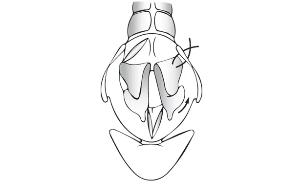
One form of a unilateral arytenoid lateralization procedure is a cricoarytenoid lateralization. This involves passing 1 or 2 sutures from the caudodorsal cricoid cartilage to the muscular process of the arytenoid cartilage. These sutures are intended to simulate the cricoarytenoideus dorsalis muscle, which is dysfunctional in dogs affected with laryngeal paralysis. Instead of a functioning muscle that allows the rima glottidis to open and close, the suture acts to rotate the arytenoid cartilage laterally, permanently enlarging the rima glottidis.
Complications
The most common complication associated with unilateral cricoarytenoid lateralization is aspiration pneumonia,3 which has been reported to occur at an incidence of 8% to 25%.2-5 After the cricoarytenoid lateralization has been performed, aspiration pneumonia may occur at any time during the dog’s life.
Other reported complications include recurrence of respiratory distress, suture failure, fragmentation of the arytenoid cartilage, coughing or gagging, and seroma formation.2-4 Bilateral arytenoid lateralization is associated with a very high complication rate and is not recommended.3
Related Article: Brachycephalic Syndrome: Innovative Surgical Techniques
Step-by-Step: Cricoarytenoid Lateralization
What You Will Need
A surgical pack
Retractor (eg, Freer periosteal elevator)
Skin hooks or stay suture
Appropriately sized (usually 2-0 to 0) nonabsorbable or slowly absorbable suture on a taper needle
A knowledgeable assistant to evaluate the rima glottidis intraoperatively
A laryngoscope and new endotracheal tube for reintubation following intraoperative evaluation or a clean location to place the existing endotracheal tube for reintubation
Additional anesthetic agent if needed for reintubation
Iris scissors or #15 scalpel blade
Gelpi retractors
Author Insight
Before the procedure, counsel the owner that the dog will likely have a voice change and is at increased risk for aspiration pneumonia. Also, the dog will likely have progressive neurological disease regardless of whether treatment is pursued.6,7

STEP 1
Position the dog in right lateral recumbency with the thoracic limbs pulled caudally. Place a roll or towel under the neck to elevate the larynx. Prepare the dog for surgery and drape in a standard fashion.
Author Insight
Spend some time with patient positioning. Placement of a roll under the neck to elevate the larynx (especially in overweight dogs) can greatly facilitate the surgical approach.
Editor’s note: This article was originally published in December 2015 as “Laryngeal Paralysis Surgery.”