Laparoscopic Liver Biopsy
Lynetta J. Freeman, DVM, MS, Diplomate ACVS, Purdue University
Laparoscopic liver biopsy is the single most requested minimally invasive procedure in our hospital. Internists rarely perform fine-needle or needle-guided biopsy procedures because these methods frequently yield inadequate samples for interpretation. Laparoscopic approaches allow visual examination of internal organs, enable multiple biopsies and retrieval of larger tissue samples from the abnormal areas, and permit visual confirmation of hemostasis without the invasiveness of open surgery.
Preoperative Concerns
Animals with hepatic disease may have coagulation defects. Preoperative platelet count, prothrombin time, and partial thromboplastin time and buccal mucosal bleeding time may help shed light on the nature of the defect. If indicated, vitamin K and fresh or frozen plasma may be given before the procedure; however, the presence of a coagulation defect is only a relative contraindication for laparoscopic liver biopsy.
Smaller incisions do not necessarily mean less risk for animals undergoing anesthesia. Laparoscopic liver biopsies are relatively short procedures, and general anesthesia is usually recommended. Airway control is important because the increased pressure from insufflation of the abdominal cavity can decrease lung volume and make it more difficult for animals to spontaneously ventilate. Monitoring pulse oximetry and end-tidal CO2 and providing assisted ventilation are recommended during these procedures.
Positioning & Preparation
The animal can be positioned in dorsal or left-lateral recumbence. Each has its advantages. The dorsal positioning allows visualization of each of the liver lobes, but is complicated by the presence of the falciform ligament, which can obscure visualization. For multifocal or systemic disease, the left lateral approach is preferred. The surgical site is prepared and draped accordingly. The Box lists necessary equipment and supplies.
Author Insight
Left lateral positioning is preferred for multifocal or systemic disease.
Step-by-Step: Laparoscopic Liver Biopsy
What You Will Need
Videolaparoscopy tower with camera, light source, insufflator, and recording device
5-mm laparoscope
2- to 5-mm trocars
5-mm endoscopic cup biopsy forceps
5-mm endoscopic blunt probe
Gelfoam absorbable gelatin sponge (pfizer.com)
Application device from a Surgitie pretied loop ligature (covidien.com)
Supplies:
Culture media
10% formalin for histopathology
Microscope slides for impression cytology
Black-topped tube for metal analysis samples

Left to right: laparoscope, 5-mm trocars, biopsy forceps, blunt probe, application device, Gelfoam absorbable gelatin sponge (inset: laparoscopic cup biopsy forceps)

Step 1: Access
Use an “open” technique to place the primary port. For animals positioned in dorsal recumbence, the initial port is placed just caudal to the umbilicus, avoiding the umbilical fat pad. If the animal is positioned in lateral recumbence, the primary port is placed at a point halfway between the caudal border of the ribs and the wing of the ilium and midway between the spine and ventral midline.
Make a 5- to 7-mm incision through the skin.
Excise the underlying subcutaneous tissue with Metzenbaum scissors down to the external fascia.
Elevate the fascia and make a small (5-mm) incision through the fascia, abdominal musculature, and peritoneum.
Confirm entry into the abdominal cavity.
Place 2 stay sutures through the abdominal fascia and use them to secure the trocar to the fascia.
Attach the insufflation tubing and insufflate to 10- to 12-mm Hg pressure with CO2.
Place a second 5-mm trocar to use for biopsy.
Step 2: Exploration
Introduce each instrument carefully into the abdominal cavity under direct visualization to avoid injury to underlying organs.
Carefully manipulate and inspect each surface of each liver lobe using the blunt probe.
Identify the area or areas you wish to biopsy. Samples are usually taken from both “normal” and “abnormal” areas of the liver. In most cases, marginal biopsy is sufficient, unless there is a focal lesion that needs to be sampled separately. Typically, samples are taken for histopathology, culture, impression cytology, and, if indicated, heavy metal analysis.
Step 3: Placement of Gelatin Sponge
For hemostasis, place Gelfoam sponge inside the abdomen before obtaining biopsy samples. To introduce the sponge, use an introducer sleeve and plastic push rod from a pretied loop ligature system (Surgitie Single-Use Ligating Loop with Delivery System). Outside the patient, make the Gelfoam sponge applier:
Break off 3 or 4 pieces of dry sponge from the sheet.
Roll sponge into 3- to 4-mm cylinders and backload into the introducer cannula (A).
Insert the plastic push rod into the cannula and the sponge applier into the working port.
Push the sponge samples into the abdominal cavity using the plastic push rod while maintaining vision with the laparoscope (B).
Position the samples near the intended biopsy site.• Remove the introducer sleeve.


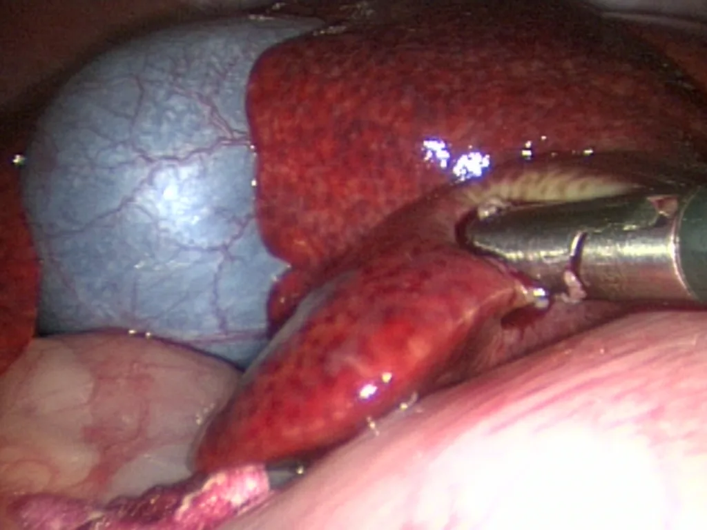
Step 4: Biopsy
Insert the laparoscopic biopsy cup forceps.
Position the forceps on tissue and close the forceps.
For a marginal biopsy, insert the closed instrument under the liver lobe next to the region to be sampled. Open the jaws and slowly withdraw the forceps while rotating the cup onto the liver margin (Figures A to D).
To sample an intrahepatic mass, press the open biopsy forceps onto the mass and apply pressure as the forceps close (Figures E and F)
Maintain the forceps in position for approximately 30 seconds.
Squeeze tighter, twist, and pull to retrieve the sample.
If necessary, slide the trocar cannula down the shaft of the biopsy forceps as it is retrieved to help in shearing the liver tissue.• Place the sample in a saline-moistened gauze sponge and pass off the surgical field.• Use the blunt probe or the biopsy forceps to “nudge” the Gelfoam samples into the tissue defect in the liver.
An alternate technique in human medicine describes use of Gelfoam plugs to occlude the biopsy tract of a 16-gauge Tru-Cut core biopsy needle in children with coagulopathy.1

A
Author Insight
Keep forceps in position for approximately 30 seconds before retrieving sample.
Step 5: Final inspection & Closure
Inspect the abdominal cavity to ensure hemostasis. If there is concern about active bleeding, it may be necessary to irrigate the site or lavage the abdomen and aspirate the fluid.
If the animal is hypotensive during surgery, bleeding can occur when the abdominal pressure is reduced and blood pressure returns to normal.
Remove each of the ancillary trocars and close each incision in the fascia, subcutaneous tissue, and skin with small sutures.
Place a lidocaine patch around each trocar site to provide local analgesia for 1 to 2 days.
Author Insight
A lidocaine patch provides analgesia for 1 to 2 days.
Author Insight
Postoperative care includes monitoring for signs of pain, bleeding, and hypoglycemia.
Postoperative Care
Most animals undergoing laparoscopic organ biopsy are ill and return to the intensive care unit for recovery and follow-up care. Patients with simple cases are sent home the day of surgery and the owners are called the following morning. Postoperative care includes monitoring for signs of pain, bleeding, and hypoglycemia. Supportive medical management is continued until a definitive diagnosis is obtained and specific therapy can be instituted.