Interpreting Dental Radiographs
Kendall Taney, DVM, DAVDC, FAVD, Center for Veterinary Dentistry & Oral Surgery, Gaithersburg, Maryland
Dental radiography should be included in a complete oral examination, as only ≈40% of the tooth and periodontium can be evaluated without radiographs. Bone loss from periodontal disease, periapical or endodontic pathology, tooth vitality, impacted teeth or retained roots, and other conditions (eg, neoplasia, tooth resorption) can also be assessed.<sup1-3 sup>
Routine full-mouth dental radiography can take minimal time when the appropriate technique is used and can be key in developing treatment plans.
Imaging Techniques
Dogs and cats have a flat hard palate; therefore, the bisecting angle and parallel techniques should be used to obtain radiographs that represent the true size of a tooth. Descriptions of these and other imaging techniques (eg, extraoral and oblique views) are available.4-7 Hands-on practice can lead to consistent results.

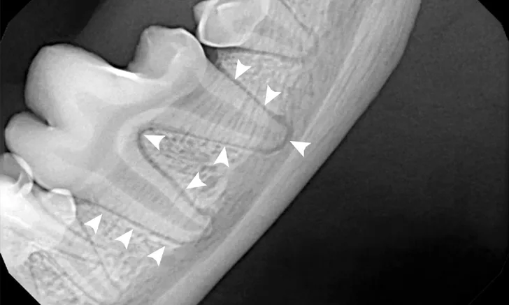
FIGURE 1A
Patient positioned in lateral recumbency; the bisecting angle technique is used to obtain radiographs of the right maxillary fourth premolar (A). The plate or film should be parallel to the hard palate (white line). The direction of the tooth root to be imaged is represented by a black line. An imaginary line can be drawn halfway between the angle of the plate or film and tooth roots (ie, bisecting angle [gray line]), usually a 45-degree angle for the maxilla. The beam (ie, x-ray tube head) should be oriented perpendicular to the bisecting angle (gray line) so the actual size of the tooth is shown (B); a crown fracture with suspected pulp exposure is visible (arrow), and radiolucency can be seen around the apex of the distal root (arrowheads).