Overview
Measurement of serum alanine aminotransferase (ALT) and aspartate aminotransferase (AST) activity is important for hepatobiliary disease screening.1-4 Serum transaminases are sensitive indicators of acute toxic, metabolic, and infectious hepatopathies and valuable in detection of subclinical stages of chronic inflammatory/necroinflammatory, fibrotic, and vascular disease. Increases in serum ALT and AST activity should not be ignored, as significant associated hepatocellular degeneration and/or necrosis are possible, and the cause should be investigated.
Epidemiology
Incidence/Prevalence
Serum chemistry profiles in dogs and cats frequently detect increases in serum ALT or AST activity. In a study that included 1,022 blood samples from both healthy and sick dogs, serum ALT and AST activity increased in 17% and 11% of dogs, respectively.5 In a separate study that included 407 healthy middle-aged dogs and 128 cats, serum ALT and AST activity were increased in 9.3% and 2% of dogs, respectively.6 Serum ALT and AST activity were both increased in 7% of cats.
Signalment
Breed Predisposition
Increases in serum ALT or AST activity may be the first indicator of congenital or acquired breed-specific diseases in dogs and cats.1-4,7,8
Examples include
Immune-mediated or copper-associated hepatitis9,10
Doberman pinschers
American and English cocker spaniels
Labrador retrievers
Bedlington terriers
Dalmatians
West Highland white terriers
English springer spaniels
Serum transaminase levels are often mildly elevated in dogs (less often in cats) with congenital vascular diseases (eg, portosystemic vascular anomalies, primary hypoplasia of the portal vein) and in dogs with disorders with distinct canine breed predispositions.8,9
Pathophysiology
ALT
In most cases, serum ALT is considered a liver-specific enzyme in dogs and cats.1-3 In dogs, severe muscle necrosis can increase serum ALT, but a muscle source for the enzyme elevation can be eliminated by simultaneous measurement of serum creatine kinase.
Elevations in serum ALT are associated with reversible or irreversible damage to the hepatocyte plasma membrane. Increased serum ALT activity has the highest sensitivity (80%-100%) for inflammation, necrosis, vacuolar hepatopathy, and primary neoplasia.2 Reduced sensitivity has been noted for the detection of liver failure caused by feline hepatic lipidosis (72%), hepatic congestion (70%), metastatic neoplasia, portosystemic vascular anomalies, and secondary hepatic lipidosis (60%).1-3
The magnitude of serum ALT elevation is generally proportional to the number of injured hepatocytes. In dogs, the elimination half-life is ≈2.5 days; a 50% decrease over 2 to 3 days is thus a good prognostic sign. In cats, the serum elimination half-life is much shorter (≈6 hours); return to normal values after an acute insult thus occurs faster.1-3
AST
Compared with serum ALT, AST is more sensitive but less specific for detection of hepatic disease.1-3 Serum AST is derived from liver, skeletal muscle, and cardiac muscle sources. In humans, there is both a cytosolic and mitochondrial liver AST isoenzyme; this is presumably also the case for dogs and cats. The cytosolic enzyme is released with reversible or irreversible damage to hepatocyte plasma membranes and usually parallels increases in serum ALT. The mitochondrial enzyme is released only with irreversible hepatocyte injury; thus, the ALT:AST ratio approaching 1 generally indicates that severe irreversible injury has occurred.7
In general, the degree of serum AST elevation for a given amount of hepatocyte damage is less than the degree of ALT elevation. If serum AST is much higher than ALT, a muscle source of the enzymes should be explored.
The elimination half-life of serum AST in dogs and cats has been reported as 12 hours and 77 minutes, respectively. Because the elimination half-life of serum AST is shorter in dogs compared with ALT, levels of the former enzyme generally return to normal faster. Hemolysis also increases serum AST.
Potential Causes of Changes in Serum ALT or AST Activity
Increases in serum transaminases are not 100% specific for primary hepatobiliary disease, as the liver can be damaged secondary to other pathologic processes occurring in the abdomen. The liver receives a rich blood supply from the arterial and portal venous systems and is a dynamic metabolic organ with various functions, including fat digestion, intermediary metabolism, and detoxification of endogenous and exogenous compounds. Extrahepatic systemic disorders and diseases in organ systems drained by the portal circulation, particularly the GI tract and pancreas, can damage the liver and increase serum ALT and AST activity. Under these conditions, increased liver enzymes may not reflect the presence of clinically important primary hepatic disease (see Conditions in Which Serum Transaminase Activity May Not Reflect Primary Hepatobiliary Disease).
Increases in serum transaminases are not always sensitive enough to pick up mild liver disease. Sensitivity depends on the nature of the disease process occurring in the liver. Serum transaminases are most sensitive for acute necroinflammatory and toxic diseases and less sensitive for chronic inflammatory/fibrotic disease, neoplasia, perfusion abnormalities, and cholestatic disorders.1-4,11,12
Some dogs, particularly Alaskan sled dogs, can have genetic mutations associated with an allele at the CFA13 locus that results in low serum ALT activity,13,14 making serum ALT elevations less likely in the presence of hepatic damage.15
Conditions in Which Serum Transaminase Activity May Not Reflect Primary Hepatobiliary Disease
Endocrinopathies
Diabetes mellitus, hyperadrenocorticism, hyperthyroidism (cats)
GI disease
Pancreatitis, inflammatory bowel disease
Neoplasia
Metastatic disease
Hypoxia/hypotension
Congestive heart failure, hypotensive crisis, severe hemolytic anemia, status epilepticus
Blunt abdominal trauma
Systemic infections
Next Steps When Evaluating Patients With Abnormal Values
The entire clinical scenario, including patient history, clinical signs, physical examination findings, diagnostic imaging, and ancillary laboratory evaluation, should be examined to determine the clinical significance of persistently elevated levels of serum ALT or AST.
The magnitude of increase in serum ALT and AST activity necessary to trigger a thorough clinical investigation has not been determined. In dogs with clinical signs, any increase in serum transaminases should be investigated. In subclinical dogs, there is some evidence that increases 4 times more than normal that last >12 weeks should trigger investigation.16 Smaller increases in subclinical patients should be rechecked in 4 to 6 weeks to determine whether elevations persist. Similar studies have not been done in cats.
Patient History
Exposure to a hepatotoxin
Cycad seeds, mushrooms, microcytins, aflatoxin, xylitol, copper
Medication or supplement known to cause drug-induced liver injury (see Hepatotoxic Drugs in Dogs & Cats)17
Note: Corticosteroids and phenobarbital are known to induce serum alkaline phosphatase in dogs but do not induce increases in serum transaminases. Increases in serum transaminases in dogs with endogenous and exogenous corticosteroid excess or in dogs receiving phenobarbital reflect the presence of hepatocellular damage.18,19
Polyuria/polydipsia
Potential causes include chronic liver disease, congenital portosystemic shunts, and hyperthyroidism (cats).
Dermatologic disease
Potential causes include hepatocutaneous syndrome.
Chronic intermittent GI signs
Potential causes include gastric ulceration due to chronic liver disease, congenital portosystemic shunts, chronic pancreatitis, inflammatory bowel disease, and hyperthyroidism (cats with palpable thyroid nodule)
Hepatotoxic Drugs in Dogs & Cats17,22,23
Acetaminophen (dogs, cats)
Amiodarone (dogs)
Anabolic steroids (dogs, cats)
Azathioprine (dogs)
Carprofen (dogs)
Chlorambucil (cats)
Corticosteroids (dogs)
Diazepam (cats)
Doxycycline (dogs)
Glucosamine/chondroitin (with manganese) (dogs)
Halothane, metophane (dogs, cats)
Ketoconazole (dogs, cats)
Leflunomide (dogs)
Lomustine (CCNU) (dogs)
Megestrol acetate (cats)
Methimazole (cats)
Methotrexate (dogs)
Phenobarbital (dogs)
Potentiated sulfonamides (dogs)
Rifampin (dogs)
Rivaroxaban (dogs)
Toceranib (dogs)
Trazodone (dogs)
Zonisamide (dogs)
Physical Examination Findings & Potential Causes
Diffuse cerebral signs
Hepatic encephalopathy due to congenital or acquired portosystemic shunts
Jaundice
Hepatobiliary disease or posthepatic obstruction
Abdominal effusion
Chronic liver disease with ascites, neoplasia, pancreatitis, congestive heart failure
Hepatomegaly
Chronic hepatitis, passive congestion, neoplasia, hepatic lipidosis (cats), amyloidosis, cirrhosis (cats)
Abdominal pain
Pancreatitis, cholecystitis, gastric ulceration
Thyroid nodule (cat)
Hyperthyroidism
Relevant Diagnostic Testing, Findings, & Potential Causes20
Diagnostic Imaging
Radiography
Hepatomegaly
Chronic hepatitis, passive congestion, neoplasia, hepatic lipidosis (cats), amyloidosis, cirrhosis (cats)
Microhepatica
Cirrhosis (dogs), congenital portosystemic shunts (dogs)
Choleliths
50% visible
Decreased abdominal detail
Ascites from portal hypertension (pure to modified transudate), pancreatitis (nonseptic exudate), bile peritonitis
Cardiomegaly with pulmonary edema/pleural effusion
Heart failure
Normal liver
Does not rule out primary hepatic disease
Hepatobiliary
Focal or multifocal lesions
Primary or metastatic hepatobiliary neoplasia, benign nodular hyperplasia (common in older dogs, typically associated with increased serum alkaline phosphatase), abscess
Diffuse hyperechoic liver
Hepatic lipidosis, vacuolar hepatopathy, lymphosarcoma, fibrosis
Diffuse hypoechoic liver
Passive congestion, lymphosarcoma, infectious hepatitis
Gallbladder/biliary tree
Gallbladder mucocele or choleliths, distended biliary tree with extrahepatic bile duct obstruction
Portal vasculature
Single congenital or multiple acquired portosystemic shunts, portal vein thrombosis, passive congestion
GI tract
Increased wall thickness, altered wall layering, dilated lacteals associated with chronic enteropathy or GI neoplasia
Pancreas
Enlarged hypoechoic surrounded by hyperechoic fat indicative of pancreatitis
Normal liver
Does not rule out primary hepatic disease
Other abdominal organs
Primary neoplasia of the spleen, stomach, pancreas, intestines, or adrenals accompanied by focal hepatic nodules
Laboratory Analysis
CBC
Serum chemistry profile
Urinalysis
Cytology of abdominal effusion
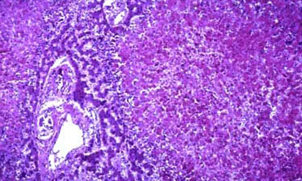
Fine-needle aspiration or liver biopsy
Determination of a primary liver disorder often relies on tissue sampling.
Neoplasia or hepatic lipidosis in cats may be diagnosed on cytology via fine-needle aspiration, but many primary disorders (particularly chronic inflammatory, fibrotic, or vascular diseases) require a histopathologic diagnosis, preferably from biopsy samples obtained via laparoscopy.21
Disease-specific testing
Primary hepatobiliary disease
Concurrent disease causing a reactive hepatopathy should be ruled out.
Hepatic function test
Total serum bile acids
Blood ammonia
Prothrombin or partial thromboplastin time
Hepatic fine-needle aspiration and/or biopsy (preferably via laparoscopy)21
Hyperthyroidism
Serum total thyroxine
Chronic GI disease
Serum B12 (ie, cobalamin) and folate
Quantitative pancreatic lipase immunoreactivity
Endoscopy with intestinal biopsy
Fine-needle aspiration or biopsy of the pancreas
Neoplasia
Biopsy or fine-needle aspiration
Disease-Specific Findings
Primary liver disease
Serum ALT and AST activity increase together, ± increases in serum alkaline phosphatase and serum gamma-glutamyl transpeptidase, hypoalbuminemia, hyperbilirubinemia, low BUN, hypoglycemia
Abnormal hepatic function tests (eg, elevated total serum bile acids, serum hyperbilirubinemia with normal packed-cell volume, blood hyperammonemia, increased prothrombin or partial thromboplastin time)
Supportive imaging findings (eg, nodularity, hepatomegaly, small liver with nodules and irregular margins, hepatic mass, gallbladder mucocele, bile duct obstruction)
Supportive findings from fine-needle aspiration and/or biopsy
Chronic liver disease
Ascites that are pure transudate with a total protein of <2.5 g/dL
Right heart failure
Ascites that are modified transudate with a total protein of >2.5 g/dL
Chronic GI disease
Panhypoproteinemia, hypocholesterolemia, lymphopenia
Increased serum amylase and/or lipase (dogs) and immunoreactive pancreatic lipase; acute nonseptic inflammation on abdominal tap
Cancer
Identification of mass lesions in the abdomen, malignant effusion on abdominal tap, high protein ascites with exfoliated cells, hemorrhagic effusion with ruptured hemangiosarcoma
Absence of neoplastic cells does not rule out a malignant effusion.
Diagnosis at a Glance
Measurement of serum transaminase levels is the most sensitive screening test available for the presence of hepatobiliary disease.
Serum ALT and AST elevations indicate active hepatobiliary disease but provide no information on the functional state of the liver.
The prognostic significance of serum ALT and AST elevations can be improved through evaluation of sequential tests, reviewing concurrent increases in other serum markers of hepatobiliary disease (eg, serum alkaline phosphatase, serum gamma-glutamyl transpeptidase, albumin, bilirubin, total serum bile acids), and using these markers in conjunction with diagnostic imaging findings ± hepatic histopathology.
Serum ALT or AST can be elevated without the presence of clinically significant liver disease, particularly in disorders that affect the GI tract and the pancreas in endocrine disorders (eg, hyperthyroidism in cats, hyperadrenocorticism in dogs).
Treatment
Treatment should be based on the cause of increased serum transaminases.
In cases of drug toxicity, the drug should be discontinued and the patient monitored for normalization of serum enzyme activity.
Prognostic Significance of Abnormal Findings
Serum transaminases correlate with the severity of hepatocellular damage but are not necessarily prognostic, as they can leak into the blood with both reversible and irreversible hepatic damage.1-3 Serum transaminase levels increase when active disease causes hepatocyte membrane damage; however, the magnitude of elevation in serum ALT or AST activity may not be indicative of the extent of functional impairment because the liver has a significant regenerative capacity and great functional reserve. Increases in serum ALT or AST activity may be mild in cases of severe, functional, end-stage disease because of enzyme depletion secondary to replacement of hepatocytes by fibrous tissue. Alternatively, in cases of acute liver failure, higher serum ALT activity is a good, not bad, prognostic indicator.11 Thus, a single serum ALT or AST measurement should never be used to establish prognosis.
The clinical significance of increased serum ALT or AST activity is improved by one or more of the following.1-3
Evaluating serum enzyme activity longitudinally for trends
Monitoring frequency should be based on the degree of elevation, whether a treatable etiology is identified, and whether the patient was ill.
Assessing for concurrent increases in other serum markers of liver disease (ie, serum alkaline phosphatase activity, serum gamma-glutamyl transpeptidase activity)
Assessing liver function (serum total bile acids) or serum bilirubin as well as BUN, glucose, and albumin levels
Obtaining a hepatic biopsy