Incisional Wedge Biopsy of Subcutaneous Tumors
Lisa Corti, DVM, DACVS, CCRP, North Shore Veterinary Surgery, Andover, Massachusetts, North Shore Community College, Danvers, Massachusetts
Tumor histopathology can determine tissue type, presence of neoplastic or inflammatory processes, cell behavior or tumor grade, appropriate staging diagnostics, treatment options, and prognosis.1 Knowledge of tissue type can also help pet owners make informed decisions.
Subcutaneous tissue tumors are common.2,3 Fine-needle aspiration can accurately differentiate neoplastic from inflammatory processes and is often the first test performed4; however, not all cytologic samples are diagnostic or accurate, especially when peripheral inflammation or necrotic centers of tumors are sampled.4,5 Surgical biopsy is therefore often pursued as it can provide tissue that reveals the underlying architecture and interplay of cells.1 Tumor grade can only be identified via biopsy.1,2,6,7
Types of Surgical Biopsy
Surgical biopsies are excisional or incisional. Excisional biopsy involves removal of an entire mass without prior knowledge of tumor type7; surgical margins may be inadequate, risking tumor regrowth, and additional aggressive surgery or adjuvant treatment (eg, radiation, chemotherapy) may be needed.7 Incisional biopsy involves excision and submission of a small piece of the tumor; this indicates the tumor type before definitive removal, and appropriate surgical margins can be planned.7
Incisional biopsies can be performed using a punch biopsy instrument, specialized biopsy needle device, or wedge technique.1,2,7 Punch biopsies of the outer layers of a subcutaneous tumor are less likely to be diagnostic because the depth of the circular blade determines the depth of the sample.7 Although needle biopsies have a high rate of diagnostic accuracy, specialized cutting needles are required and advanced imaging modalities may be needed to increase the accuracy of sample retrieval.1,2,6,7 Wedge biopsy can provide direct visualization of abnormal tissue, control of the length and depth of the sample, and avoidance of specialized equipment use.1,7
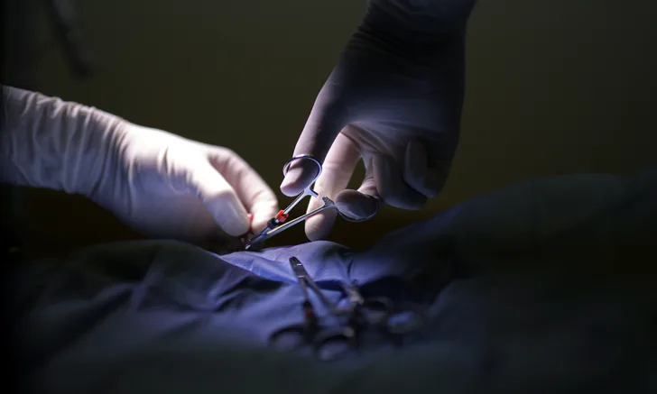
Anesthesia is not always needed for wedge biopsies; heavy sedation, analgesics, and a local anesthetic are often sufficient.

Large left ischial and proximal thigh mass in a dog with ulceration and tumor rupture through the skin. This ulcerated area should be avoided during wedge biopsy.
Biopsy Location
Sample location is an important consideration for wedge biopsy of a subcutaneous tumor. Sampling from the junction of a lesion and normal tissue is commonly recommended for dermatologic biopsies3,7; however, sampling a subcutaneous mass at this junction can lead to inadvertent spread of cancer cells beyond the original tumor.1,7 Removal of the original biopsy site should be considered if definitive tumor excision is performed later, as the biopsy site is considered contaminated with tumor cells. Sampling closer to the center of the mass may be preferable, but areas of inflammation or necrotic debris should be avoided (Figure 1).
Understanding the appearance of healthy tissue and normal tissue layering is key to distinguishing neoplastic from normal tissue. Familiarity with regional anatomy overlying and adjacent to a subcutaneous mass can help avoid inadvertent damage to vital structures (eg, blood vessels, nerves; Figure 2). Many tumors have a capsule or pseudocapsule, which should be grossly differentiated from normal tissue layers and penetrated during dissection to obtain a sample of the underlying tumor.2,7
Narrow and deep wedge biopsies are most likely to obtain an accurate sample.7 Because the wedge biopsy site is considered contaminated, and a lengthy incision that may contain cancer cells can make definitive removal with adequate margins challenging, the skin incision should not span the entire width of the tumor. Depending on the size of the mass, the incision can vary from a few millimeters to a few centimeters.


Small subcutaneous mass over the distal radius and carpus in a dog. Awareness of the cephalic and accessory cephalic vein (A; solid arrow) locations can help avoid inadvertent laceration of these vessels. Incision was made through the subcutaneous tissue lateral to the vessel (B; dashed arrow).
Sample Submission
The pathology form should be filled out completely when a biopsy sample is submitted so the pathologist can provide an informed, accurate, and clinically relevant diagnosis (see Pathology Form).8 A study found >88% of biopsy submission forms supplied inadequate clinical information in at least one key content area, and 3% of forms were devoid of information beyond signalment.9 Inadequate information significantly influenced reporting of clinical information by the pathologist.9
Pathology Form8
A pathology form should include:
Signalment
Diagnostic information and pertinent results from blood work, hormonal tests, radiography, ultrasonography, and advanced imaging (eg, MRI, CT)
Tumor site (specific), duration, and rate of growth
Gross description of the tumor, including size, shape, character, associated tissue layer, vascularity, and ulceration
Tumor-associated clinical signs (eg, lameness, pruritus, skin changes)
Type of biopsy (eg, incisional, excisional) and method used (eg, punch, needle, wedge)
Previous tumor treatments
Results of previous tumor-related tests (eg, cytology, biopsies) and diagnostics (eg, radiography, ultrasonography, CT)
Step-by-Step: Incisional Wedge Biopsy
What You Will Need
Clippers
Sterile surgical scrub and alcohol
Sterile gown and gloves
Sterile instrument pack
Scalpel blade handle
Brown-Adson thumb forceps
Needle holders
Suture scissors
#10 or #15 scalpel blade
Small biopsy jar with 10% buffered formalin
3-0 synthetic absorbable monofilament suture (eg, poliglecaprone, polydioxanone)
3-0 synthetic nonabsorbable monofilament suture (eg, nylon)
Histopathology form

Step 1
Clip and aseptically prepare the mass and surrounding skin.

Step 2
Apply sterile drapes around the tumor or biopsy site.
Author Insight
Ulcerated areas should be draped out of the field of view when possible.
Step 3
Incise through the skin (A) and underlying subcutaneous tissue (B) over the mass.


Author Insight
Length of the incision should not span the entire mass.
Step 4
Identify and incise the tumor capsule (A, arrow; subcutaneous lipoma) or pseudocapsule (B, dashed arrow; subcutaneous sarcoma).2,7


Author Insight
A true tumor capsule and a tumor pseudocapsule are distinguished histologically; grossly, they appear similar. The tumor capsule is usually found deep to the subcutaneous fat and can be separated from the adipose layer for better identification. Dissection of highly vascular, friable, or amorphous tumors may not be possible.
Step 5
Incise the tumor, slicing down and inward as if following the side of an inverted triangle (A). Remove the scalpel blade and angle it in the opposite direction, slicing down and inward on the other side of the imaginary inverted triangle (B).


Author Insight
Depth of the inverted triangle should be greater than the width of its base.7 A nondiagnostic biopsy result is commonly caused by sampling only the superficial tissues surrounding a mass. It is important to cut into the tumor. When uncertain on how deep to make the wedge, it is better to cut deeper.
Step 6
Continue incising into the tumor until the point of the inverted triangle is reached and the wedge is released. Place the specimen in 10% neutral buffered formalin immediately after excision to limit tissue artifacts.8

Author Insight
The ratio of tissue to 10% neutral buffered formalin should be 1:10.8
Tumor sample shown here has 2 distinct regions (solid arrow, circular and homogeneous; dashed arrow, layered and heterogeneous). Gross and microscopic variations in the same sample can help provide a more accurate histopathologic diagnosis.8
Step 7
Close the subcutaneous and subcuticular layers and the skin layers routinely.

Author Insight
Hemorrhage is often a concern, and certain tumor types (eg, hemangiosarcoma, highly vascular soft tissue sarcomas) can bleed considerably. This should not deter incising deeply into the tumor. Hemorrhage can be controlled by closing the biopsy site with a cruciate or horizontal mattress suture pattern across the junction of normal and abnormal tissue. Direct digital pressure or hemostatic powder and foam can be used if the tissue is friable. If a blood vessel is inadvertently lacerated, it can be clamped with a mosquito hemostat and ligated or cauterized. Applying ice to the incision during recovery and at home can help limit bleeding and hematoma formation.