Inapparent Oronasal Fistula
Profile
Definition
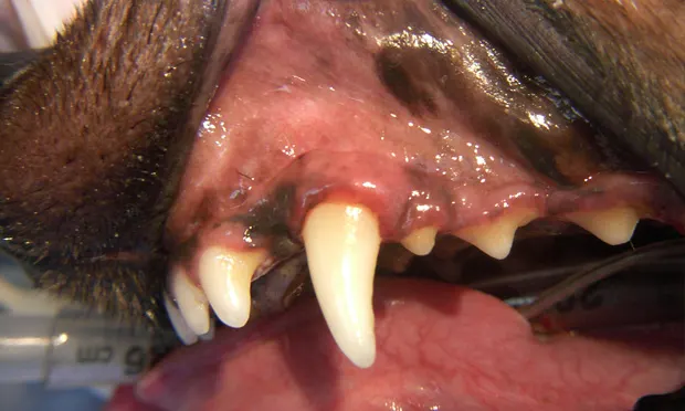
An opening from the mouth to the nasal cavity that occurs adjacent to teeth that appear clinically healthy (Figure 1).1-3
Figure 1. (above) Eight-year-old, spayed female Dachshund with normal-appearing gingiva over the buccal aspect of the maxillary left canine tooth (#204). This patient has a history of chronic sneezing. The tooth appears normal visually. Without probing, the problem would be missed.
Signalment
Can develop in any breed dog; extremely common in dachshunds; also common in poodles, schnauzers, Yorkshire terriers, and breeds prone to periodontal disease.3
Common on the palatal aspect of the maxillary canine tooth, but also found on the palatal aspect of some incisor and premolar teeth.
Many cases are bilateral.
Typically occurs in older pets with periodontal disease, but as the name suggests, many of these maxillary canine teeth appear normal clinically.
Causes
Most cases are caused by periodontal tissue destruction on the palatal aspect of the maxillary canine teeth (see pathophysiology section).
Some cases are caused by trauma from malocclusions, such as occlusion of the tip of lingually displaced mandibular canine teeth, especially when the mandible is shorter than the maxilla.
Some cases are caused by trauma, such as luxation or avulsion of the maxillary canine.
Some cases are iatrogenic.
Pathophysiology
The periodontal tissues (gingival attachment, periodontal ligament, cementum, and alveolar bone) are destroyed on the mesial, palatal, and distal aspects of maxillary teeth, most often the maxillary canine tooth. Periodontal disease can be insidious.
Plaque bacteria cause some tissue destruction through the release of cytotoxic enzymes.
The host immune response to the plaque bacteria is also responsible for some periodontal tissue destruction by enzymes and chemotactic factors.
The periodontal disease occurs primarily on the palatal aspect of the maxillary canine teeth in an area not easily seen or accessible for routine home care; the result is periodontal tissue destruction along the palatal aspect of the maxillary canine teeth.
The roots of maxillary teeth extend adjacent to the nasal cavity, and the destruction of the soft tissue and bone between the roots and nasal cavity results in deep periodontal pockets or an opening/fistula.
The chronic influx of saliva, food, water, and oral bacteria results in chronic rhinitis.
Recent research has identified specific black pigmented anaerobic bacteria associated with some alveolar bone loss (see Treatment, Prevention).
Signs
Chronic rhinitis
Unilateral or bilateral nasal discharge: serous, purulent, bloody, or combination
Sneezing
Possibly, clinical signs of periodontal disease such as plaque, gingivitis, and calculus
Clinical malocclusions causing traumatic oronasal fistula
Pain Index
Pain may be nonexistent to moderate.
Many patients appear to have no sensitivity, but some are reluctant to have pressure applied over the dorsal nasal cavity or alveolar juga.
If debris has accumulated in the nasal cavity, then discomfort may be an issue.
Diagnosis
Definitive Diagnosis
Usually made with a periodontal probe to measure pocket depths in the area of the suspected disease (Figure 2).
Sterile saline flushed into periodontal pockets while the body and base of the head is elevated and the nose tipped down may reveal fluid from one or both nares.
With probing/flushing of deep periodontal pockets, care must be taken not to create iatrogenic oronasal fistula. If a fistula is found when using mild to moderate pressure, it would have been unlikely that fistula formation could have been prevented.
Periodontal probing depths should be recorded; if no fistula is noted, then the periodontal disease should be treated (see Treatment) to salvage this tooth or prevent chronic disease.

Figure 2. Periodontal probing on the palatal aspect of the left maxillary canine tooth (#204) may reveal a deep periodontal pocket.
Other Diagnostic Testing
Dental radiography is imperative to diagnose most periodontal disease, but because of the location of the alveolar bone loss and the positioning of radiography of the maxillary canine tooth, dental radiographs usually don't show the true extent of disease. Radiographs may reveal alveolar bone loss on the mesial and distal aspects of the maxillary canine teeth. Radiographs may be more valuable for showing alveolar bone loss in premolar teeth with associated oronasal fistulas.
Rhinoscopy may reveal granulation tissue or areas of inflammation at the level of the oronasal fistula.
Nasal biopsy specimens of these areas show nonspecific inflammation.
CT may be of value to show bone loss in the future, but expense is the limiting factor.
Differential Diagnosis
Nasal foreign body
Fungal rhinitis
Nasal neoplasia
Bacterial or viral rhinitis
Treatment
Deep Periodontal Pockets
Periodontal pockets that have not yet broken through to fistula formation (Figures 3 and 4)4
Periodontal pocket less than 5 mm: Closed root planing with a hand curette or appropriate subgingival ultrasonic or sonic scaler and subgingival curettage (removal of the granulation tissue and diseased gingival tissue within the periodontal pocket-also called periodontal pocket debridement).
Periodontal pockets greater than 5 mm: Surgical elevation of a gingival flap and open root planing with a hand curette or appropriate ultrasonic or sonic scaler and debridement of diseased gingival tissue (subgingival curettage).
Guided tissue rejuvenation: Placement of a perioceutic (Doxirobe; Pfizer Animal Health, www.pfizer.com) in periodontal pockets greater than 4 mm will help reduce further collagen breakdown and provide local antibiotic therapy for healing tissue. A very good first step to do 2 to 4 weeks before guided tissue regeneration.
To regain lost alveolar bone in this area, in many cases, bulk osseous implants, such as Consil (Nutramax Laboratories, www.nutramaxlabs.com), or osseoinductive materials, such as PepGen P-15 (DENTSPLY; Friadent Ceramed, www.ceramed.com), can be used to form bone.
Prevention: Recent research has shown potential benefit of using a killed bacterin against the Porphyromonas bacteria responsible for a substantial amount of alveolar bone loss. Pfizer has received conditional license from the USDA for a vaccine, Porphyromonas denticanis, gulae, salivosa Bacterin vaccine. It targets the 3 species that have been shown to be most pathogenic in dogs. While the approval is for use in healthy dogs, the vaccine may help patients with familial tendencies toward inapparent oronasal fistula formation or those that have been treated with guided tissue regeneration to prevent subsequent bone loss.
Treatment Once Fistula Has Formed
The only proven method to resolve an oronasal fistula is extraction of the involved tooth or teeth and closure of the fistula via a mucoperiosteal flap (Figures 5-8).2,3,5
Basic oral surgical principles are important:
Maintain blood supply.
Make gingival flaps larger than defects to cover.
Eliminate tension on the gingival flap when the defect is covered.
Suture fresh-cut mucosa to fresh-cut mucosa (this is an important point when closing inapparent oronasal fistulas because the intact epithelial edges of the defect will inhibit proper healing if not trimmed, resulting in closure failure).
Place sutures over connective tissue, in this case bone, whenever possible.
Use multiple flaps (double-flap closure) when single-flap closures have failed. If the previous oral surgical principles are maintained, most oronasal fistula repairs should be successful without the need for double-flap closures.
Minimize the use of cautery.
Anecdotal reports have described placing a collagen base layer (Curasorb, Tyco Healthcare/Kendall, www.tycohealthcare.com) deep in oronasal fistulas, followed by placement of osseoinductive material (PepGen P-15) to induce regeneration of bone in the oronasal defect. It is important to point out that no known published studies indicate that this approach is effective.
Client Education
These lesions can be bilateral.
Chronic rhinitis and chronic infection may have systemic implications.
Following surgical extraction and oronasal fistula closure, expect to see bloody or blood-tinged nasal discharge for 24 to 48 hours. Recommend that the owners confine patient for 2 to 4 days to a quiet place that is easily cleaned if the patient sneezes.
Soft diet only for 4 weeks.
No chew toys for 4 weeks.
Reevaluation at 2 and 4 weeks (sedate and probe if necessary).
Hospitalize for 24 to 48 hours if necessary.
Follow-up
Patient Monitoring
Immediately after procedure, expect some bloody nasal discharge with or without sneezing.
For deep periodontal pockets treated as outlined above: reevaluation with periodontal probing under sedation at 6 weeks, 3 months, 6 months, and 1 year. Abnormal probing depths may necessitate retreatment or extraction.
For surgical extraction and closure of fistulas: if healed at the 4-week recheck, these should stay that way unless traumatized. If healed at the 4-week recheck, patient can resume normal eating and chewing activities.
In General
Relative Cost
Prevention is based on early diagnosis and homecare $-$$$
Treatment can be costly for anesthesia and repair $$$$-$$$$$
Prognosis
Prognosis for complete recovery is very good to excellent. The effect of the elimination of chronic nasal infection is not measurable but should have systemic benefits.